Download
1 / 15
150 likes | 267 Views
Mechanics of Ventilation. Prof. K. Sivapalan. Introduction. Gas exchange occurs in alveoli Atmospheric air must go to alveoli and expelled after exchange. Negative pressure is created in the lungs for sucking air by the muscles.

E N D
Mechanics of Ventilation Prof. K. Sivapalan
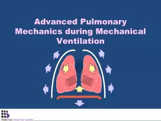
Introduction • Gas exchange occurs in alveoli • Atmospheric air must go to alveoli and expelled after exchange. • Negative pressure is created in the lungs for sucking air by the muscles. • The lung recoils like a balloon and expels the air in it- elastic fibers. Mechanics of Ventilation
Pleural Cavity. • The lungs are separated from the chest wall and the diaphragm by the pleural cavity containing a thin film of fluid. • The surface tension allows sliding but keeps both surfaces attached to each other. • Expansion- chest muscles out words and the diaphragm downwards. Mechanics of Ventilation
Respiratory Muscles • Diaphragm – contraction pulls the lung downwards. • Increase diameter of the chest cavity- • Elastic fibers in chest wall • External intercostals • Sternocleinomastoid • Anterior serrati • Scalini • Decreasing the chest cavity- • Elastic recoil of lungs • Internal intercostals • Abdominal muscles Mechanics of Ventilation
Changes in Pressure and Volume in Quiet Inspiration • Diaphragm and external intercostals contract. • Elasticity of the wall facilitates but the elasticity of lungs and surface tension in alveoli opposes • Pleural pressure decreases to -7.5 cm H2O • Intrapulmonary pressure falls to -1 • Air flows in. Mechanics of Ventilation
Changes in Pressure and Volume in Quiet Expiration • Diaphrgm and external intercostals relax. • Pleural pressure increases to -5 cm H2O – not to zero. • Elasticity of the wall is over powered by the elasticity of lungs and surface tension in alveoli. • Intrapulmonary pressure rises to +1 • Air flows out Mechanics of Ventilation
Surface Tension in Alveoli • The surface tension in alveoli tends to collapse the alveoli. • Surfactant secreted by type II alveolar epithelial cell reduces the surface tension from 72 dynes/cm to 5-30. • Surfactant is a mixture of protein, phospholipids and ions. • Respiratory distress syndrome- failure of alveoli to open in premature babies – secretion of surfactant after 7 months or later. Mechanics of Ventilation
Pleural pressure • The pleural cavity is under negative pressure during quite breathing. • Forced inspiration creates more negative pressure. • Positive pressure is observed in forced expiration, cough and sneeze. • Any communication with atmosphere either through lung or chest wall will suck air and results in Pneumothorax- lung collapses and chest wall expands. • Closed, open and tension pneumathorax. • Loss of elasticity of lungs- barrel shaped chest Mechanics of Ventilation
Lung Volumes • Lung volumes are measured by a spirometer. • Tidal volume- inspired or expired with normal breath- 500 ml in adult male • Inspiratory Reserve Volume- extra volume inspired by maximal inspiration. [3 L] • Inspiratory Capacity = TV+IRV • Expiratory Reserve volume- extra volume expired by maximal expiration.[1.1 L] • Residual Volume- Volume remaining after maximal expiration.[1.2 L] • Functional Residual Capacity= RV+ERV • Vital Capacity=ERV+TV+IRV- maximal expiration after maximal inspiration. Mechanics of Ventilation
Forced Vital Capacity • FVC is the Vital capacity obtained by forced expiration after maximal inspiration. • FEV1 is the fraction of the FVC expelled in the FIRST SECOND.[>80%]. Also peak flow. • MVV- maximal voluntary ventilation- 125-170 L/m Mechanics of Ventilation
Dead Space • Dead space is the non functional – not participating in gas exchange- space in the respiratory tract • Anatomical- nose, pharynx, trachea, ..up to terminal bronchiole. • Equals in ml to approximately the weight in pounds [150 ml] • Physiological- includes nonfunctional alveoli as well Mechanics of Ventilation
Ventilation • Minute ventilation = • average TV x average RR • 500x12= 6000 ml/minute • Alveolar Ventilation- volume of air moved into alveoli per unit time • [TV-DV]x RR = [500-150]x12 = 4200 ml/min • Effects of increasing and decreasing dead space • Ventilation to different alveoli depends on gravity and expansion Mechanics of Ventilation
Perfusion • Volume of blood flowing through the lungs per minute [5 L /min at rest] • Perfusion of different area within the lung is influenced by gravity. • When erect, more blood flows to the base. • Lying causes venous congestion Mechanics of Ventilation
Features of Pulmonary Circulation • Pulmonary artery ressure- 25/8 mmHg. • Arterioles constrict in reduced oxygen and increased carbondioxide. • Pulmonary capillary pressure is 7 mmHg. • Pulmonary oedema is accumulation of fluid in alveoli. Mechanics of Ventilation
Ventilation Perfusion Ratio • Ratio between [alveolar] ventilation and perfusion- V/P = 4.2/5.5 =0.8 • The V/P of local areas in the lung vary due to gravity and disease. • If not compensated adequately- defective gas exchange. Mechanics of Ventilation