Download
1 / 15
161 likes | 444 Views
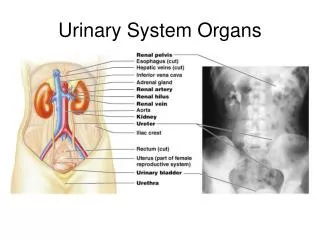
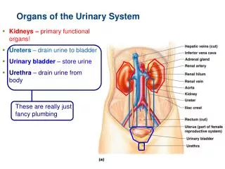
URINARY SYSTEM ORGANS. Kidneys (2) Ureters (2) Urinary bladder Urethra. kidneys. Kidneys are paired organs that lie on the posterior abdominal wall on either side of the vertebral column. The right kidney is slightly left than the lower kidney

E N D
URINARY SYSTEM ORGANS • Kidneys (2) • Ureters (2) • Urinary bladder • Urethra
kidneys • Kidneys are paired organs that lie on the posterior abdominal wall on either side of the vertebral column. • The right kidney is slightly left than the lower kidney • On medial concave boarder of each kidney is the hilus • The hilus transmits the renal pelvis, the renal artery, the renal vein and the sympathetic nerve fibers
coverings • Fibrous capsule: closely applied to te outer surface • Perirenal fat: covers the fibrous capsule • Renal fascia: it encloses the kidney and the suprarenal fat. • Pararenal fat: external to the renal fascia
Renal structures • The renal parenchyma is divided into an outer cortex and inner medulla • The medulla is composed of approximately 12 renal pyramids, each having its base oriented toward the cortex and its apex (renal papilla) projecting medially. • The cortex extends into the medulla between adjacent pyramids as the renal columns • Extending from the bases of the renal pyramids into the cortex are striations called medullary rays.
Within the renal sinus, the upper expanded end of the ureter divides into 2 or 3 major calyces, each of which in turn divides into 2 or 3 minor calyces .
Ureters • The ureters are muscular tubes leading from the renal pelvis to the lower bladder. • Each ureter measures approximately 10 in. in length and has an upper expanded end called renal pelvis.
Ureteric constrictions • Where the renal pelvis joins the ureter • Where it is kinked as it crosses the pelvic brim • Where it pierces the bladder wall
Urinary bladder • It is located immediately behind thee pubic bones within the pelvis. • The bladder has a maximum capacity of~ 500 ml. • The empty bladder is pyramidal in shape • When the bladder fills it bcomes ovoid in shape and superior surface rises in the abdomen. • The muscle coat (detrusor muscle) consists of 3 layers of smooth muscle fibers.
Urethra • 3 – 4 cm long in females • Bound by connective tissue to anterior wall of vagina • Urethral orifice exits body between vaginal orifice and clitoris
Urethra • ~18 cm long in males • Prostatic urethra • ~2.5 cm long, urinary bladder prostate • Membranous urethra • ~0.5 cm, passes through floor of pelvic cavity • Penile urethra • ~15 cm long, passes through penis
The male urethra has three regions: • prostatic urethra • 2) membranous urethra • 3) penile urethra. Difficulty in voiding urine with enlarged prostate