Download

1 / 33
330 likes | 781 Views
Developments in Non-Invasive Whole-Body Molecular Tumor imaging. Lu Yin (Lucy). Outline. Molecular imaging Whole body photonic tumor imaging. Radiolabelled peptides in molecular imaging Cell penetrating peptide in whole body tumor imaging. Molecular imaging.

E N D
Developments in Non-Invasive Whole-Body Molecular Tumor imaging Lu Yin (Lucy)
Outline • Molecular imaging • Whole body photonic tumor imaging. • Radiolabelled peptides in molecular imaging • Cell penetrating peptide in whole body tumor imaging
Molecular imaging • Molecular imaging, which I define here as the “noninvasive, quantitative, and repetitive imaging of targeted macromolecules and biological processes in living organisms,” requires two basic elements: (i) molecular probes whose concentration and/or spectral properties are altered by the specific biological process under investigation and (ii) a means by which to monitor these probes. ---------Harvey R. Herschman Science. 2003 Oct 24;302(5645):605-8. www.medical.philips.com/.../mi_home.jpg
Imaging probes • Light- or near-infrared (NIR) emitting molecules Radioisotopes labeled molecules Detections: Photonic imaging Positron emission tomography (PET) Single photon emission computed tomography (SPECT) Magnetic resonance imaging (MRI) X-ray computed tomography (CT) • Direct binding ----- monitor receptor e.g. (3-(2-[18F]fluoroethyl)spiperone([18F]FESP)}used to monitor the dopamine receptors of the striatum Indirect binding ----- monitor the activity of their targets e.g. hexokinase substrate 2-deoxy-2-[18F]fluoro-Ddeoxyglucose (FDG), which monitors glucose metabolism .
Whole-body photonic imaging of small animal • Small animal imaging is an important translation tool between in vitro research and clinical application. Whole-body photonic imaging • Planar imaging (muti-photon or confocal microscopy) The simplest technique for detecting optical reporter molecules in vivo, uses photographic principles to capture light emitted from the animals----fluorescence reflectance imaging (FRI). Limitations: Signal projection view Limited penetration depth (<1mm) Obscured signal due to tissue heterogeneity limited spatial resolution • Tomography imaging Fluorescence molecular tomography (FMT) Bioluminescence tomography Photoacoustic tomography
Spectral imaging applied to in vivo fluorescence detection (a) Standard color image obtained from a green fluorescence protein (GFP)-expressing mouse implanted with a red fluorescent protein (RFP)-expressing tumor. (b) Spectral imaging and processing improves visualization of the RFP signal, which can be separated from the mouse intrinsic auto-fluorescence and GFP fluorescence.
Planar imaging VS Tomography imaging Planar imaing
Performance characteristics of Planar and Tomography imaging • FMT gives better resolution with depth • FMT gives better signal with varying background • FMT is able to image fluorochromes in highly diffusive medium that simulates tissue optical heterogeneity.
Improving spatial sampling Image reconstruction is based on mathematical models that describe the composite photon propagation in tissue and in air. The animal surface was captured using photo-grammetry, that is, the mathematical combination of photographs obtained under different angles to deduce the physical dimension of the animal.
Visualization of brain structure and function using photoacoustic tomography (PAT) PAT also referred to as optoacoustic or thermoacoustic tomography, attains the advantage of combining ultrasonic-scale spatial resolution with high sensitivity to tissue light absorption and can yield information on physiology or on exogenously administered light absorbers. (a,b) Functional maps of brain activities corresponding to the left-side (a) and right-side (b) whisker stimulations, respectively, acquired with the skin and skull intact.
Radiolabeled peptide in oncology Weiner RE, Thakur ML. BioDrugs. 2005;19(3):145-63.
Radionuclide imaging of small-cell lung cancer (SCLC) using 99mTc-labeled neurotensin peptide 8–13 • Neurotensin (NT) is a 13 amino acid peptide originally isolated from calf hypothalamus. • It has dual function of neurotransmitter or neuromodulator in the nervous system and of local hormone in the periphery. neuromedin N neurotensin • Three subtypes of neurotensin receptors; two of them belong to the family of G protein-coupled receptors, whereas the third one is an entirely new type of neuropeptide receptor and is identical to • gp95/sortilin, a 100 kDa-protein with a single transmembrane domain. Neurotensin (NT) receptors are overexpressed in a variety of tumors e.g. prostatic tumors and pancreatic tumor. Jean-Pierre Vincent, et al. TiPS – July 1999 (Vol. 20) Kaijun Zhang, et al. Nuclear Medicine and Biology 33 (2006) 505–512
Synthesis of (Na-His)Ac-NT(8–13) derivative NT(8–13), the C-terminal hexapeptide fragment (Arg8–Arg9–Pro10–Tyr11–Ile12–Leu13) of NT, contains the amino acids essential for binding NT receptors Redio-labeled with [99mTc(H2O)3(CO)3]+
Cell-penetrating peptides (CPPs) • The first CPP came from the discovery that the third helix of Antennapedia homeodomain, pAntp(43–58), can cross biological membranes. • Different CPPs have been described to efficiently deliver various types of cargo to the inside of cells,from low molecular weight drugs to liposomes, plasmids, antibodies or nanoparticles. • CPPs are peptides made of less than 30 amino acids that are internalised via energy-dependent or independent mechanisms. Positively charged amino acids, hydrophobicity and amphipathicity are common features shared among many of the known CPPs. http://hugin.ethz.ch/wuthrich/people/billeter/antphd.jpg http://homeobox.biosci.ki.se/homeo/antp1.gif Sílvia Pujals et al. Biochimica et Biophysica Acta 1758 (2006) 264–279
Internalization mechanisms Self-assembly The exact internalization macheniss are still not clear yet.Initially, CPPs were defined as short cationic peptides able to translocate through the plasma membrane of eukaryotic cells via a receptor- and endocytosis-independent mechanism, but a re-evaluation of the internalisation mechanism yielded a new CPP concept. Endocytosis . Sílvia Pujals et al. Biochimica et Biophysica Acta 1758 (2006) 264–279
Principal classes of CPPs Sílvia Pujals et al. Biochimica et Biophysica Acta 1758 (2006) 264–279
Tumor imaging by means of proteolytic activation of cell-penetrating peptides Fig. 1. Schematic diagram of activatable CPPs. Cellular uptake induced by a cationic peptide is blocked by a short stretch of acidic residues attached by a cleavable linker. Once the linker is cleaved, the acidic inhibitory domain drifts away, and the cationic CPP is free to carry its cargo into cells.
How to target tumor? • MMP-2 (matrix metalloproteinase ) Cleavable linkerPLGLAG • MMPs Matrix metalloproteinases (MMPs) are a large family of zinc metallo-endopeptidases that degrade varying components of the extracellular matrix in both normal and diseased tissue. More Than 22 members, all of which share a common catalytic core containing a zinc molecule in the active site. Aberrant MMP activity is believed to be associated with a number of pathological conditions including rheumatoid arthritis, tumor invasion, and metastasis. MMP-2 and MMP-9, have been widely studied in breast and prostatic tumors, the growth of which is often hormone-dependent.
ACPPs Adopt a Hairpin Conformation Before Cleavage Nuclear Overhauser effects observed in two-dimensional NMR of a simple ACPP, succinyl-e8-XPLGLAG-r9-Xk, where X denotes 6-aminohexanoyl. Dashed red lines indicate observed nuclear Overhauser effects, and the green line highlights the peptide outline for clarity.
Until Cleaved Off, Polyanionic Sequences Inhibit Association of CPPs with Cells
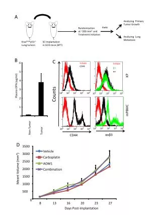
MMP-2 Cleavable ACPPs Concentrate in Human Tumors Xenografted into Mice quantitation (fluorescence intensity of tumor - autofluorescence)/(fluorescence of normal contralateral region - autofluorescence) mean ± SE ACPP 2.1 ± 0.17 n = 6 scrambled isomer 1.3 ± 0.16, n = 2 all-D-amino acid 1.5 ± 0.11, n = 4 HT-1080 tumor xenografts visualized with activatable CPPs. HT-1080 tumors were implanted into the mammary fat pad of nude mice and allowed to grow until they reached 5-7 mm in diameter. (A1) A live anesthetized animal imaged 50 min after injection with 6 nmol of cleavable peptide. (A2 and A3) Tumor and muscle histology from a different animal killed 30 min after injection. (B1-B3) A similar experiment with the scrambled peptide. (Scale bars, 30 μm.)
Quantitation of Tissue Distribution by Cy5 fluorescence moles injected into animal/total body weight moles of recovered peptide/weight of tissue sample SUV
ACPPs Light Up Human Squamous Cell Carcinomas ACPP staining of surgically resected human squamous cell carcinoma tissue. Fresh tumor tissue was sliced in 1-mm slices and incubated in 1 μM cleavable (A) or uncleavable (B) peptide for 15 min, washed, and frozen. Sections (5 μm) were taken for fluorescence microscopy by using a 10× objective, and tissue type was verified by hematoxylin/eosin stain. (A) The arrow indicates a differentiated keratin pearl. As a control, histologically normal tissue from the same patient was treated similarly with MMP-2 cleavable peptide (C) or scrambled peptide (D). (C) The arrow indicates a ring of invading tumor cells.
Contrast, (tumor tissue fluorescence - autofluorescence)/(normal tissue fluorescence - autofluorescence),was almost eight in this example. • Contrast tended to be greatest where the tumor tissue had a high histologic grade of malignancy. • lymphocytic granulation tissue was nearly as bright as the tumors themselves, possibly because of the release of MMPs from lymphocytes. • Normal tissue immediately adjacent to tumor tissue was noticeably brighter than more remote normal tissue, possibly because of the presence of immune cells or to diffusion of the soluble proteases.
Conclusion • For whole-body photonic imaging, tomography imaging gives better results compared with planar imaging. PAT will be the possible future direction of photonic imaging. Tumor imaging largely rely on probes that can target the tumor: • The 99mTc-labeled neurotensin peptide 8–13 could image the small-cell lung cancer (SCLC) by targeting overexpressed neutotesin recpetor. • MMP-2 proteolytic activation of cell-penetrating peptides can target the MMP-2 positive tumor without target specific receptor. It is a novel method that can be applied to tumor imaging.