Understanding the Cerebellum: Configurations, Functions, and Connections
The cerebellum, located in the posterior cranial fossa, plays a critical role in movement coordination, balance, and cognitive functions. Composed of two hemispheres connected by the vermis and divided into lobes, it features distinct structures like Purkinje cells and various afferent and efferent connections. The superior, middle, and inferior cerebellar peduncles facilitate communication with the brainstem and other areas. This intricate system is vital for maintaining posture, equilibrium, and muscle tone, alongside aiding motor learning and planning refined limb movements.

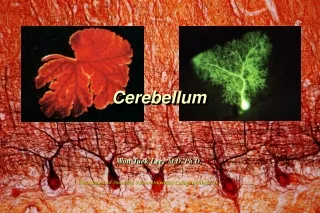
Understanding the Cerebellum: Configurations, Functions, and Connections
E N D
Presentation Transcript
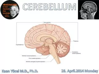
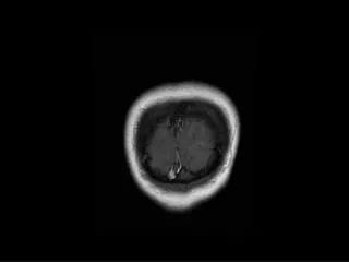
Cerebellum External Configurations - located in posterior cranial fossa - tentorium cerebelli (cerebrum), 4th ventricle (brain stem) - communicate with other structure via superior, middle, and inferior cerebellar peduncle - longitudinal division Vermis, Paravermal Region, Cerebellar Hemisphere - transverse division Anterior Lobe ------------ primary fissure Posterior Lobe ------------ posterolateral fissure Flocculonodular Lobe
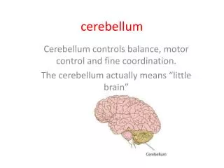
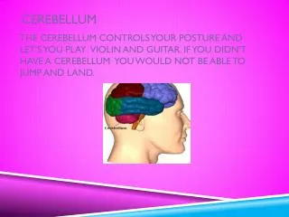
F. The cerebellum- Has two lobed hemispheres that connect via the vermis. It has outer gray matter, imbedded white matter and deep gray matter (nuclei). Its surface ridges are called folia and they are separated by fissures. It coordinates movement and makes it smooth, it helps in maintaining posture and equilibrium and is involved in cognition.
Spinocerebellum Pontocerebellum Vestibulocerebellum
1. Purkinje cell 2. granule cell 3. basket cell 4. Golgi cell 5. stellate cell 6. climbing fiber 7. mossy fiber 8. parallel fiber 9. inferior olivary nucleus 10. deep cerebellar nuclei
Cerebellum Connections Afferent Connections (1):
Vestibulocerebellar fiber linkage inferior cerebellar peduncle flocculonodular lobe vestibular nuclei medical longitudinal fasciculus motor neuron of extraocular muscles vestibulospinal tract motor neuron of trunk muscle • maintain the body’s balance and regulate ocular movement.
Superior Cerebellar Peduncle Anterior Spinocerebellar Tract Cerulocerebellar fiber Raphecerebellar fiber Rubrocerebellar fiber Hypothalamocerebellar fiber
Spinicerebellar fibra links and function direct cerebellar tract inferior cerebellar peduncle superior cerebellar peduncle vermis vestibular nucleus reticular formation of brain stem vestibulospinal tract fastigial nucleus reticulospinal tract intermedial nucleus red nucleus of opposite side rubrospinal tract lateralcorticospinal tract cortex of cerebellar hemisphere zona rolandica control muscular tension and regulate muscular movement
The fibra links and function of cerebrocerebellum pontine nucleus lateral part cortex of cerebellar hemisphere extensive area of cerebral cortex ventrolateral nucleus of dorsal thalamus zona rolandica dentate nucleus rubrospinal tract red nucleus lateral corticospinal tract • dominate the planning and coordination of refined movement of limbs
Cerebellum Connections Efferent Connections : 1. Superior Cerebellar Peduncle Cerebellothalamic fiber - from 3 deep nuclei to VPLo, VLc, CL Cerebellorubral fiber - from nucleus interpositus and dentate nucleus 2. Inferior Cerebellar Peduncle Fastigiovestibular fiber
Two types of fibers to cerebellar cortex 1. Climbing fibers a, project to dendritic trees of Purkinje neurons b. are extremely excitatory c. primarily originate from inferior olivary nucleus may be some contribution from pontine and reticular nuclei 2. Mossy fibers a. much more numerous than climbing fibers b. origination is spinocerebellar and pontocerebellar c. project to granule cell dendrites d. from granule cells, information is passed to dendritic branches of Purkinje cells. e. are extremely excitatory
Climbing and Mossy fibers are part of a circuit 1. excitatory input enters, is integrated throughout cells of cerebellar cortex, exits via axons of Purkinje cells 2. outflow is inhibitory 3. primary target of cerebellar outflow is deep cerebellar nuclei a. globose and emboliform axons project to red nucleus & reticular formation via superior cerebellar peduncles, then to cord via rubrospinal tract. b. dentate axons project to thalamus via superior cerebellar peduncles, and then to cerebral cortex c. fastigial axons project to vestibular nuclei and nuclei for cranial nerves III, IV, and VI via inferior cerebellar peduncles
CerebellumFunction Maintenance of Equilibrium - balance, posture, eye movement Coordination of half-automatic movement of walking and posture maintenace - posture, gait Adjustment of Muscle Tone Motor Leaning – Motor Skills Cognitive Function
Motor Skill Pablo Casals
Posture Gait – Ataxia Tremor
a b c Cerebellar Ataxia Ataxic gait and position: Left cerebellar tumor a. Sways to the right in standing position b. Steady on the right leg c. Unsteady on the left leg d. ataxic gait d
Cerebellar Medulloblastoma Cerebellar tumors on vermis - Truncal Ataxia - Frequent Falling The child in this picture: - would not try to stand unsupported - would not let go of the bed rail if she was stood on the floor.