acute stroke
Syahrul Department of Neurology Faculty of Medicine, Syiah Kuala University Banda Aceh, March 29, 2011. acute stroke. T he third leading cause of death T he leading cause of serious, long-term disability Indonesia : Riskesdas Depkes RI, 2007 Prevalence of stroke 8,3 per 1.000 people

acute stroke
E N D
Presentation Transcript
Syahrul Department of Neurology Faculty of Medicine, Syiah Kuala University Banda Aceh, March 29, 2011 acute stroke
The third leading cause of death • The leading cause of serious, long-term disability • Indonesia : Riskesdas Depkes RI, 2007 • Prevalence of stroke 8,3 per 1.000 people • Mortality : stroke 15,4%, hypertensive 6,8% & ischemic heart disease 5,1% • Stroke Statistics,U.S. Statistics,2010 • 143,579 people die each year from stroke • Each year, about 795,000 people suffer a stroke • About 600,000 of these are first attacks, and • 185,000 are recurrent attacks STROKE
A major economic burden on healthcare system Incidence is expected to increase 25% by 2050 Ischemic stroke, when arteries are blocked by blood clots (emboli) or by the gradual build-up of plaque other fatty deposits. (Approximately 80% of stroke are ischemic) Hemorrhagic stroke, occur when a blood brain breaks leaking blood into the bain. (20% of all stroke) Stroke
Patologi Anatomi • Stroke Iskemik • Trombosis Serebri • Emboli Serebri • Stroke Hemoragik • Perdarahan Intra Serebral • Perdarahan Sub-Arakhnoid • Perjalanan Klinis • Transient Ischemic Attack • Reversible Ischemic Neurological Defisit • Stroke In-evolution • Komplit Stroke • Sirkulasi Serebral • Stroke Sirkulasi Serebral Anterior • Stroke Sirkulasi Serebral Posterior Klasifikasi
Approach : • Pathophysiology • Clinical Signs & Symptoms • Diagnostic Supports • Neuro-Pharmacology Intervention Recent Mangementof acute ischemic stroke
Ischemic core and penumbra in human stroke (Stroke. 1999;30:93-99)
Ischemic core and penumbra in human stroke (Stroke. 1999;30:93-99)
Cellular Injury During IschemiaConsequences of Calcium Overload
Cellular Injury During IschemiaCellular Changes During Ischemia
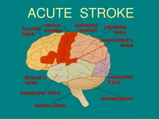
Clinical Signs & Symptoms Anatomy of Stroke
MRI : Brain Gold Standard Coronal orientation: in a slice dividing the head into front and back halves.Sagittal orientation: in a slice dividing the head into left and right halves.Axial orientation: in a slice dividing the head into upper and lower halves.
MRIin Acute Ischemic Stroke Left: diffusion-weighted MRI in acute ischemic stroke performed 35 minutes after symptom onset. Right: apparent diffusion coefficient (adc) map obtained from the same patient at the same time.
MRIin Acute Ischemic Stroke Diffusion-perfusion mismatch in acute ischemic stroke. The perfusion abnormality (right) is larger than the diffusion abnormality (left), indicating the ischemic penumbra, which is at risk of infarction.
MRIin Acute Ischemic Stroke Left: Perfusion-weighted MRI of a patient who presented 1 hour after onset of stroke symptoms. Right: Mean transfer time (MTT) map of the same patient.
Cerebral Angiography(Cerebral Angiogram, Cerebral Arteriogram, Digital Subtraction Angiography [DSA])
Echocardiogram Examines the heart through the chest (called transthoracicechocardiogram, or TTE), and one that examines the heart through the throat (called transesophagealechocardiogram, or TEE)
Atrial fibrilation • CAD, Ischemic heart disease • Infarct myocard (acute, acute) • RBB, LBB • LVH, RVH T inversion; Q pathology; ST depretson; ST elevation Electrocardiogram(EKG, ECG)
Blood routine, Glucose, Lipid Profile, Uric Acid Fibrinogen, Agregation of Trombocyte,INR Protein C, S; Anticardiolipin Antibody (ACA) Laboratory test
Optimization of medical treatment is key in the care of the stroke patient and we should be cautious when prognosticating early in the setting of acute stroke and be aware of the potential effect ‘do not resuscitate’ status may have on patient outcome J NeuroIntervent Surg 2011;3:34-37 Neurocritical Care Intervention
Prehospital Management Hospital Management Emergency Medical Service Facilities for Emergency Stroke Care “Time is brain”
Medical emergency, early hospital management Time depedent therapy Rapid confirmation (CT scan or MRI) Urgent investigation (cause of stroke) Acute therapy Comprehensive risk factor management (antihypertensive therapy, early rehabilitation, discharge planning) “Time is brain”
rt-PA Intravenous Recombinant Tissue Plasminogen Activator The ‘’engine for emergency stroke” Beneficial within 3 hours of stroke onset (NINDS 1995, PROACT II study 1999, National Stroke Foundation 2007, AHA/ASA 2007) World Stroke Congress, Seoul Korea, 20104 hours Trombolysis
Antithrombotic Therapy After the onset of stroke (>3 hours) aspirin 325 mg Anticoagulant Therapy After the onset of stroke (emboli )(3 – 8 hours) Antithrombotic & Anticoagulant
The acute treatment window for ischemic stroke is the loading of aspirin and clopidogrel within 36 hours of symptom onset of stroke Treated with 325 mg of aspirin and 375 mg of clopidogrel within 36 hours of symptom onset Loading with 375 mg of clopidogrel and 325 mg of aspirin appears to be safe when administered up to 36 hours after stroke and transient ischemic attack onset in this pilot study. Neurologic deterioration may be decreased and warrants further study . J Stroke Cerebrovasc Dis. 2008; 17(1): 26–29. Aspirin andClopidogrelin the Acute Treatment of Ischemic Stroke
When and how to treat hypertension in acute ischemic stroke? The effect of BP modification during the acute phase of ischemic stroke on functional outcome isstrongly dependent on age. (Hypertension2009; 54:769-774) When & How To Treat Hypertension ?
Loss of CBF Regulation During Acute Ischemic Stroke Hypertension 2009;54;702-703
Autoregulation of cerebral blood flow in a normal brain and in the ischemic penumbra (the tissues surrounding the ischemic core after a stroke) In the normal brain, cerebral blood flow is kept at 50 mL/100 g per minute, despite continuous fluctuations of mean blood pressure between 70 and 120 mm Hg (continuous line). Any increase in pressure leads to vasoconstriction and any decrease to vasodilation, which prevents the risk of cerebral hyper- and hypoperfusion, respectively. Above and below the limits of cerebral blood flow autoregulation, cerebral perfusion passively follows the perfusion pressure. In the ischemic penumbra, tissue perfusion follows perfusion pressure (dashed line): any fall in blood pressure may precipitate ischemia, while an increase in blood pressure may cause edema and hemorrhagic transformation. CMAJ, March 1, 2005; 172 (5)
Mostly as mono-therapywas common among a history of hypertension Angiotensin-converting enzyme inhibitors (ACEI) 65 (45.6%) Diuretics 41 (34.5%) ACEI were used in combination with diuretics in 29 (23.4%) In Cochrane review found no evidence that giving calcium antagonists after an ischemic stroke saves lives or prevents disabilities. Anti-hypertensive Medicationsin the Acute Ischemic Stroke
Recent Advances in the Treatment of Hypertensive Emergencies Crit Care Nurse 2010;30: 24-30
Rapid onset of action Predictable dose response Titratable to desired BP Minimal dosage adjustment Minimal adverse effects Easy conversion to oral agents Acceptable cost-to-benefit ratio Does not impair blood flow to vital organs (No sudden dips in BP; Does not decrease cardiac output) Does not increase ICP Normalizes CBF autoregulatory curve The Ideal Acute Antihypertensive Agent
Prevention of Early Ischemic Injury • N-Methyl-D-Aspartate Receptor Antagonists • Modulation of Non-NMDA Receptors • Nalmefene • Lubeluzole • Clomethiazole Free Radical Scavengers and Trapping Agents NXY-059 • Prevention of Reperfusion Injury • Antiadhesion Antibodies • Membrane Stabilization • Neuronal Healing Neuroprotective Agents in Stroke