Download
1 / 27

E N D
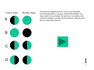
Linkes Auge Rechtes Auge A B C D Versuchen Sie anhand der Folie „Arrow“ die folgenden Gesichtsfeldausfälle zu erklären. Welche Hirnstruktur, bzw. Auge wurde wie geschädigt? Es hilft sich vorzustellen, daß man Nervenbahnen zerschneiden könnte/kann. (Dunkel stellt den Gesichtsfeldausfall dar.) 1
Their receptive field looks like this: Erklären Sie in Worten weshalb das Modell unten Orientierungsselektivität im Cortex erklären kann. Was ist eigentlich Orientierungsselektivität. Was ist ein On- oder Off-subfeld? (siehe Folie „Arrow“ aber auch spätere Vorlesungen) Wie sehen die rezeptiven Felder in der Retina bzw. im CGL (LGN) aus (Arrow). Weshalb können dann daraus (durch welche Verschaltung?) cortikale Felder entstehen? 2
Erklären Sie nebenstehendes Diagram in Worten (Arrow). Wenn man mit eine Elektrode nicht exakt vertikal in den Cortex einsticht, dann mißt man eine sich mit der Elektrodenposition ändernde Orientientierungsselektivität. Ändert sich diese immer kontinuierlich? Was ist ein Vortex? (siehe hier „map“ Vorlesung) 3
The Interdisciplinary Nature of Computational Neuroscience What is computational neuroscience ?
Different Approaches towards Brain and Behavior Neuroscience: Behavior Reaction Environment Stimulus
Black Box Psychophysics (human behavioral studies): Environment Stimulus Behavior Reaction
Neurophysiology: Behavior Reaction Environment Stimulus
Theoretical/Computational Neuroscience: Behavior Reaction Environment Stimulus
1m CNS 10cm Sub-Systems 1cm Areas / „Maps“ 1mm Local Networks 100mm Neurons 1mm Synapses 0.1mm Molecules Levels of information processing in the nervous system
CNS (Central Nervous System): CNS Systems Areas Local Nets Neurons Synapses Molekules
Cortex: CNS Systems Areas Local Nets Neurons Synapses Molekules
CNS The Phrenologists view at the brain (18th-19th centrury) Systems Areas Local Nets Neurons Synapses Molekules Where are things happening in the brain. Is the information represented locally ?
Results from human surgery CNS Systems Areas Local Nets Neurons Synapses Molekules
Results from imaging techniques – There are maps in the brain CNS Systems Areas Local Nets Neurons Synapses Molekules
Visual System: More than 40 areas ! Parallel processing of „pixels“ and image parts Hierarchical Analysis of increasingly complex information Many lateral and feedback connections CNS Systems Areas Local Nets Neurons Synapses Molekules
Primary visual Cortex: CNS Systems Areas Local Nets Neurons Synapses Molekules
CNS Systems Areas Local Nets Neurons Synapses Molekules V1 contains a retinotopic map of the visual Field. Adjacent Neurons represent adjacent regions in the retina. That particular small retinal region from which a single neuron receives its input is called the receptive field of this neuron. Retinotopic Maps in V1: V1 receives information from both eyes. Alternating regions in V1 (Ocular Dominanz Columns) receive (predominantely) Input from either the left or the right eye. Each location in the cortex represents a different part of the visual scene through the activity of many neurons. Different neurons encode different aspects of the image. For example, orientation of edges, color, motion speed and direction, etc. V1 decomposes an image into these components.
Their receptive field looks like this: CNS Systems Areas Local Nets Neurons Synapses Molekules Orientation selectivity in V1: Orientation selective neurons in V1 change their activity (i.e., their frequency for generating action potentials) depending on the orientation of a light bar projected onto the receptive Field. These Neurons, thus, represent the orientation of lines oder edges in the image. stimulus
CNS Systems Areas Local Nets Neurons Synapses Molekules Thus, neurons in V1 are orientation selective. They are, however, also selective for retinal position and ocular dominance as well as for color and motion. These are called „features“. The neurons are therefore akin to „feature-detectors“. Superpositioning of maps in V1: For each of these parameter there exists a topographic map. These maps co-exist and are superimposed onto each other. In this way at every location in the cortex one finds a neuron which encodes a certain „feature“. This principle is called „full coverage“.
Orientation selective cortical simple cell CNS Systems Areas Local Nets Neurons Synapses Molekules Local Circuits in V1: stimulus Selectivity is generated by specific connections
Layers in the Cortex: CNS Systems Areas Local Nets Neurons Synapses Molekules
CNS Systems Areas Local Nets Neurons Synapses Molekules Local Circuits in V1: Circuit LGN inputs Cell types Spiny stellate cell Smooth stellate cell
At least all these things need to be considered when making a „complete“ cortex model Considerations for a Cortex Model • Input • Structure of the visual pathway • Anatomy of the Cortex • Cell Types • Connections • Topography of the Cortex • „X-Y Pixel-Space“ and its distortion • Ocularity-Map • Orientation-Map • Color • Functional Connectivity of the cortex • Connection Weights • Physiological charateristics of the neurons
CNS Systems Areas Local Nets Neurons Synapses Molekules Structure of a Neuron: At the dendrite the incoming signals arrive (incoming currents) At the soma current are finally integrated. At the axon hillock action potential are generated if the potential crosses the membrane threshold The axon transmits (transports) the action potential to distant sites At the synapses are the outgoing signals transmitted onto the dendrites of the target neurons
Different Types of Neurons: dendrite dendrite Bipolar cell Unipolar cell axon soma axon soma Retinal bipolar cell (Invertebrate N.) Different Types of Multi-polar Cells Hippocampal pyramidal cell Purkinje cell of the cerebellum Spinal motoneuron
rest Ion channels: Cell membrane: Cl- K+ Membrane - Circuit diagram: