Download
1 / 1
10 likes | 23 Views
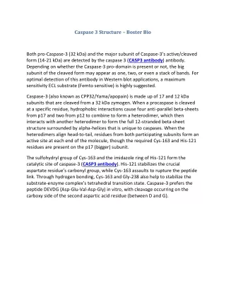
Both pro-Caspase-3 (32 kDa) and the major subunit of Caspase-3u2019s active/cleaved form (14-21 kDa) are detected by the caspase 3 (CASP3 antibody) antibody.

E N D
Caspase 3 Structure – Boster Bio Both pro-Caspase-3 (32 kDa) and the major subunit of Caspase-3’s active/cleaved form (14-21 kDa) are detected by the caspase 3 (CASP3 antibody) antibody. Depending on whether the Caspase-3 pro-domain is present or not, the big subunit of the cleaved form may appear as one, two, or even a stack of bands. For optimal detection of this antibody in Western blot applications, a maximum sensitivity ECL substrate (Femto sensitive) is highly suggested. Caspase-3 (also known as CPP32/Yama/apopain) is made up of 17 and 12 kDa subunits that are cleaved from a 32 kDa zymogen. When a procaspase is cleaved at a specific residue, hydrophobic interactions cause four anti-parallel beta-sheets from p17 and two from p12 to combine to form a heterodimer, which then interacts with another heterodimer to form the full 12-stranded beta-sheet structure surrounded by alpha-helices that is unique to caspases. When the heterodimers align head-to-tail, residues from both participating subunits form an active site at each end of the molecule, though the required Cys-163 and His-121 residues are present on the p17 (bigger) subunit. The sulfohydryl group of Cys-163 and the imidazole ring of His-121 form the catalytic site of caspase-3 (CASP3 antibody). His-121 stabilizes the crucial aspartate residue’s carbonyl group, while Cys-163 assaults to rupture the peptide link. Through hydrogen bonding, Cys-163 and Gly-238 also help to stabilize the substrate-enzyme complex’s tetrahedral transition state. Caspase-3 prefers the peptide DEVDG (Asp-Glu-Val-Asp-Gly) in vitro, with cleavage occurring on the carboxy side of the second aspartic acid residue (between D and G).