Animal Development
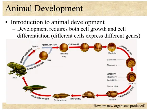
Animal Development. Ch 47 Fertilization through organogenesis. Stages of Human Development. Fertilization Zona pellucida First cell division Cleavage Blastomere Holoblastic cleavage Meroblastic cleavage regulation Morophogenesis Gastrulation Organogenesis. Fertilization.

Animal Development
E N D
Presentation Transcript
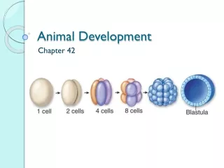
Animal Development Ch 47 Fertilization through organogenesis
Stages of Human Development • Fertilization • Zonapellucida • First cell division • Cleavage • Blastomere • Holoblastic cleavage • Meroblastic cleavage • regulation • Morophogenesis • Gastrulation • Organogenesis
Fertilization • Female secretions increase sperm motility and change structure to cause fertilization potential (capacitation) • Moist environment necessary for sperm • First six hours
Fertilization • Zonapellucida contain receptor cite and acrosomal reaction which binds sperm to egg • Changes cause slow polyspermy to prevent additional sperm from entering egg • No fast polyspermy reactions in mammals
Figure 47.5 Zona pellucida Follicle cell Corticalgranules Spermnucleus Spermbasal body
Fertilization • First Cell Division • Mitosis forms true nuclei in daughter cells • 12-36 hours after sperm bonding • Each cell is now a blastomere • http://www.hhmi.org/biointeractive/human-embryonic-HHMI embryonic development
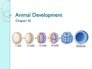
Cleavage • Rapid cell division with almost continuous S and M phases of cell cycle • Little or no protein synthesis (G1 or G2) • Blastula- Hollow ball of cells form with blastocoel cavity
Figure 47.6 50 m (d) Later blastula (a) Fertilized egg (c) Early blastula (b) Four-cell stage
Cleavage • In frogs and mammals is holoblastic • “holo” means complete • Humans have 3 divisions in first three days with little yolk forming • Birds and reptiles cleavage is meroblastic (incomplete) to get extensive yolk formation • The “ends” of the blastula are called the animal pole and vegetal pole • Gray crescent is the area on the opposite side from sperm binding
Figure 47.7 Zygote 2-cellstageforming Gray crescent 0.25 mm 8-cell stage (viewedfrom the animal pole) 4-cellstageforming Animalpole 8-cellstage 0.25 mm Blastula (at least 128 cells) Vegetal pole Blastocoel Blastula(crosssection)
Regulation of Cleavage • The total mass of the structure does not change from zygote to blastula, the cells just get smaller • Cells divide until the ratio of material in each nucleus to cytoplasm is sufficiently large • Small cells balance the amount of DNA to mRNA for protein synthesis (think surface are to volume ratio)
Morphogenesis • Transformation of embryo orientation and shape • Important is the cell shape, position and survival • Two important phases: • Gastrulation- establishment of cell layers • Organogenesis- formation of organs
Morphogenesis:Gastrulation • During gastrulation there is a mass movement of cells which results in the blastula becoming a gastrula • Three germ layers develop • ectoderm- outside layer • mesoderm- middle layer • endoderm- inside layer • Some organisms (cniderians) do not have a mesoderm • HHMI Differentiation and Cell Fate • http://www.hhmi.org/biointeractive/differentiation-and-fate-cells
Figure 47.8 ECTODERM (outer layer of embryo) • Epidermis of skin and its derivatives (including sweat glands, hair follicles) • Nervous and sensory systems • Pituitary gland, adrenal medulla • Jaws and teeth • Germ cells MESODERM (middle layer of embryo) • Skeletal and muscular systems • Circulatory and lymphatic systems • Excretory and reproductive systems (except germ cells) • Dermis of skin • Adrenal cortex ENDODERM (inner layer of embryo) • Epithelial lining of digestive tract and associated organs (liver, pancreas) • Epithelial lining of respiratory, excretory, and reproductive tracts and ducts • Thymus, thyroid, and parathyroid glands
Animalpole Figure 47.9 Blastocoel Mesenchymecells Gastrulation in Sea Urchin Vegetal plate Vegetalpole Blastocoel Filopodia Mesenchymecells Archenteron Blastopore 50 m Blastocoel Ectoderm Archenteron Blastopore Key Mouth Future ectoderm Mesenchyme(mesoderm formsfuture skeleton) Digestive tube (endoderm) Future mesoderm Anus (from blastopore) Future endoderm
2 3 1 CROSS SECTION Figure 47.10 SURFACE VIEW Animal pole Blastocoel Gastrulation in Frog Dorsal lip ofblasto-pore Dorsal lip ofblastopore Blastopore Earlygastrula Vegetal pole Blastocoelshrinking Archenteron Ectoderm Blastocoelremnant Mesoderm Endoderm Key Future ectoderm Blastopore Lategastrula Future mesoderm Yolk plug Archenteron Blastopore Future endoderm
Fertilized egg Figure 47.11 Primitivestreak Gastrulation in Chick Embryo Yolk Primitive streak Epiblast Future ectoderm Blastocoel Endoderm Migratingcells(mesoderm) Hypoblast YOLK
1 3 4 2 Endometrial epithelium(uterine lining) Blastocyst reaches uterus. Figure 47.12 Gastrulation in Human Inner cell mass Uterus Trophoblast Blastocoel Blastocyst implants(7 days after fertilization). Expanding region oftrophoblast Maternal bloodvessel Epiblast Hypoblast Trophoblast Expanding region oftrophoblast Extraembryonic membranesstart to form (10–11 days),and gastrulation begins(13 days). Amniotic cavity Epiblast Hypoblast Yolk sac (from hypoblast) Extraembryonic mesoderm cells(from epiblast) Chorion (from trophoblast) Gastrulation has produced athree-layered embryo withfour extraembryonicmembranes. Amnion Chorion Ectoderm Mesoderm Endoderm Yolk sac Extraembryonic mesoderm Allantois
Morphogenesis:Gastrulation in Humans • 1. Blastocyst first 6 days • Fertilization occurs in the oviduct • Inner cell mass becomes the embryo which is the source for stem cells • Little yolk (stored nutrients) Inner cell mass Blastocyst reaches uterus.
Morphogenesis:Gastrulation in Humans • 2. Trophoblast 7 days after fertilization • Outer epithelium secretes enzymes for implantation which allows for blood to surround trophoblast • Epiblast-upper layer becomes the embryo • Hypoblast- lower layer Maternal bloodvessel
Morphogenesis:Gastrulation in Humans • 3. Extraembryonic membranes 10-11 days • Formed by embryo • Enclose special structures outside the embryo • Gastrulation begins day 13 when implantation is complete • Cell migration occurs as cells move inward from epiblast through primitive streak to form mesoderm and endoderm Chick gastrulation
3 Figure 47.12c Expanding region oftrophoblast Amniotic cavity Epiblast Hypoblast Yolk sac (from hypoblast) Extraembryonic mesoderm cells (from epiblast) Chorion (from trophoblast) Extraembryonic membranesstart to form (10–11 days),and gastrulation begins(13 days).
Morphogenesis:Gastrulation in Humans • 4. End of gastrulation • Three germ layers are formed • Extraembryonic layers from placenta • These layers are an evolutionary necessity in land (dry) environments
4 Figure 47.12d Amnion Chorion Ectoderm Mesoderm Endoderm Yolk sac Extraembryonic mesoderm Allantois Gastrulation has produced athree-layered embryo withfour extraembryonicmembranes.
Organogenesis • More localized changes HHMI