Download
1 / 35
470 likes | 601 Views
Learn about the key methods for analyzing solid samples, including structural properties and surface phenomena, using cutting-edge technologies such as X-ray diffraction and mass spectrometry. Discover the importance of characterization in understanding material compositions and behaviors.

E N D
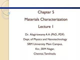
Chapter 5Materials CharacterizationLecture 1 Dr. Alagiriswamy A A (PhD., PDF) Dept. of Physics and Nanotechnology SRM University Main Campus, Ktr., SRM Nagar, Chennai, Tamilnadu
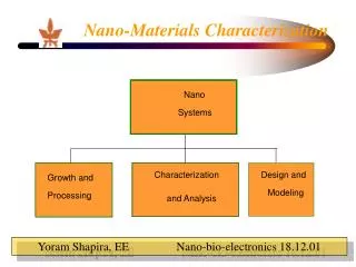
Characterization of Solid Samples Structural Properties Surface phenomena Bulk elemental composition IR spectrom. Dynamic SIMS. Dissolution step Direct solid analysis UV-Vis spectrom Static SIMS Atomic absorption Electron. microscope XRD/FESEM SEM/FESEM TEM./HRTEM ICP spectrometry SEM-EDX XPS/SAM ICP mass spect. CHN analyser dilatometry. AFM Excitation Source Detector Sample
, Heisenberg uncertainty principle limits simultaneous knowledge of conjugate Variables. This is because of intrinsic nature
Scattering by One Electron Oscillation direction of the electron The scattered energy (intensity) is: X-ray beam Electron Electric vector of The incident beam
Scattering by Two Electrons p q s0 2 r 1 s 2 1
Single molecule diffraction? • The scattering by a single electron is weak, but not too weak: • A crystal amplifies “the” signal by summing up the amplitudes from ~1024 electrons; Don’t you even need crystals rather than looking at single electron diffraction pattern????
Neutron diffraction • = h/p; = 2.860 / E1/2 • Fast moving neutrons, slow, thermal,resonance neutrons, cold, • Atomic nuclei could scatter neutrons, and emit beta particles
X-ray Fluorescence (XRF) Methods Schematic diagram of an X-ray fluorescence system utilizing an analyzing crystal
XRF - Features Energy dispersive X-ray fluorescence technology (ED-XRF) provides one of the simplest, most accurate and most economic analytical methods for the determination of the chemical composition of many types of materials. It is non-destructive and reliable, requires no, or very little, sample preparation and is suitable for solid, liquid and powdered samples. It can be used for a wide range of elements, from sodium (11) to uranium (92), and provides detection limits at the sub-ppm level; it can also measure concentrations of up to 100% easily and simultaneously
X-ray Attenuation characteristic TARGET X-ray (Compton) (Rayleigh) • The basics of XRF are very similar to those of EPMA—we are dealing with characteristic x-rays and continuum x-rays— with the exception that we are doing secondary fluorescence : x-ray spectroscopy of our samples using x-rays coming out of a sealed tube to excite the atoms in our specimen. • The big difference is that • there is NO continuum generated in the sample (x-rays can’t generate the Bremsstrahlung), and • we are using BOTH characteristic x-rays of the sealed tube target (e.g., Cr, Cu, Mo, Rh) AND continuum x-rays to generate the characteristic x-rays of the atoms in the sample. • XRF has been a bulk analytical tool (grind up 50-100 grams of your rock or sample to analyze), though recently people are developing “micro XRF” to focus the beam on a ~100 mm spot.
Si(Li) Detector FET Window Super-Cooled Cryostat Dewar filled with LN2 Si(Li) crystal Pre-Amplifier Cooling: LN2 or Peltier Window: Beryllium or Polymer Counts Rates: 3,000 – 50,000 cps Resolution: 120-170 eV at Mn K-alpha
Quantitative Analysis Concentration Intensity