Download
1 / 65
650 likes | 692 Views
Explore the epidemiology of head injury, causes of head trauma, brain injury types, anatomic features, characterization, and treatments like concussion, contusion, laceration, countercoup injury, and diffuse axonal injury.

E N D
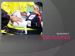
Epidemiology of Head Injury • Epidemic in all countries • In USA; *2.000.000 medically evaluated patients for year *400.000 hositalization *75.000 deaths
Leading cause of death between ages 15-40 • Major cause of perminant disability
Head Trauma What are the causes of head trauma? *motor vehicle accidents(%50) *Falls(%21) *Violence(%12) *Home, work and sporte accidents(%17)
Brain Injury after head trauma 1. Primary injury (At the time of Impact) Primary brain injury refers to the sudden and profound injury tothe brain that is complete at the time of impact. This occurs at the time of the car accident, gunshot wound, or fall. 2. Secondary brain injury Secondary brain injury refers to the changes that evolve over a period of time (from hours to days) after the primary brain injury. It includes an entire cascade of cellular, chemical, tissue, or blood vessel changes in the brain that contribute to further destruction of brain tissue.
Characterization of Traumatic Brain Injury • Anatomic features(general) -Blunt vs penetrating -closed(acceleration-deceleration) vs open(open scalp and skull) -focal(for example contusion, hemorrhage) vs. diffuse(focal lesions produce dysfunction specific to disturbed area;diffuse lesions are associated with disturbance of consciousness, cognitive or memory problems) -hemorrhagic vs non hemorrhagic
*Anatomic features(specific) -No radiographic abnormality -Skull fracture (linear, compound, depressed, diastatic, skull base fractures) -Epidural hematoma -Subdural hematoma -Cerebral contusion/intracerebral hemorrhage -Subarachnoid hemorrhage -Diffuse injury(tissue shear)
A) There are types of brain injury that can occur at the time of impact These are; Concussion Contusion Laceration Countercoup Injury Diffuse Axonal Injury
1) Concussion • Brief LOC with normal exam, normal CT scan • Patients may have mild lethargy or confusion • Treatment: observation • Avoid reinjury: contact sports are excluded until all symptoms and signs are fully recovered • Post concussion syndrome:headaches, depression and personality changes
2) Cerebral Contusion • Area of focal injury with deficit upon area injured (heterogenous hemorhagic areas, edema, brain necrosis) • Often occur after coup- counter coup pattern(e.g. frontal and occipital) • % 20 of contusions expand into the surgical hematoma • Observe patient in the ICU, repeat CT scan within 24 hours. Monitor ICP unless patient is conscious and cooperative.
3) Laceration *tears in brain tissue caused by a foreign object or pushed in bone fragment from a skull fracture 4) Countercoup injury *As the brain jolts backwards, it can hit the skull on the opposite side and cause a bruise called a counter coup lesion.
5) Diffuse Axonal Injury • High speed injury with streching or shearing of the brain tissue(usually caused by acceleration/deceleration head traumas) • Often cited as the cause of loss of consciousness in comatose patients without a lesion on CT. • Cerebral edema and ICP elevation is common • Mortality 30-40%, good outcome 20-30%
B) Scalp Laceration/ Avulsion • Most common head injury after head trauma. • Due to its vascularity, it bleeds a lot. • Generally does not cause hypovolemia in adults, but may be a reason in children.
C) Skull Fractures Skull fractures may cause underlying brain and dura damage, or not 1) Linear Fractures (most common): *Usually not identified in field (suspect based on mechanism of injury and overlying soft tissue swelling) *Usually not emergent.
2)Depressed Fracture:Bone segment is pushed inward and can cause brain damage (neurologic signs and symptoms are evident); needs surgical repair. • 3) DiastaticFracture:Fracture line transverses one or more sutures of the skull causing a widening of the skull. It usually occurs in infants and young children; but may also occur in adults (esp. The lambdoidal suture is affected).
4)BasilarFracture:A basilar skull fracture (or basal skull fracture) is a fracture of the base of the skull, typically involving the temporal bone, occipital bone, sphenoid bone, and/or ethmoid bone. • This type of fracture is rare, occurring as the only fracture in just 4% of severe head injury patients. • Common cause of death in auto racing accidents.
Basilar fractures are difficult to detect on X-rays. Generally diagnosis is made clinically by finding: *CSF otorrhea *CSF rhinorrhea *Periorbital ecchymosis(Raccoon eyes) *Battle’s sign:ecchymosis of the mastoid processes of the temporal bone
Nondisplaced fractures usually heal on their own; but if there is a damage at cranial nerves, if there is CSF otorrhea or rhinorrhea or a displaced fracture, special attention is required.
5)Open Skull Fractures: *Cranial contents are exposed *We should manage like evisceration *If possible protect the exposed tissues with a moist, clean dressing and operate as soon as possible.
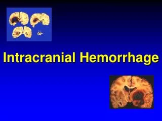
D)Intracranial Hemorrhages 1)Epidural Hematomas 2)Subdural Hematomas 3)Subarachnoidal Hemorrhages
Intracranial Hemorrhage Early S/S Severe head pains Dizziness Nausea Unequal pupil sizes Sleepiness Severe S/S Deteriorating consciousness Neck rigidity Slow pulse Slow respiration Convulsions
1) Epidural Hematoma %0.5 of all head traumas, %9 of all comatous head traumas Classically arterial (%70) (MMA), %30 venous (MMV or dural sinus) 70-80% occur with temporoparietal fracture & middlemeningeal artery rupture Blood accumulates between external periosteal layer of dura and calvaria
Epidural Hematoma “Classic” presentation (10-33% of cases) brief LOC followed by lucid interval of several hours Amnesia Contralateral hemiparesy Ipsilateral pupillary dilatation (Hutchinson’s pupil) Obtundation, severe headache, vomiting, focal neurologic signs May progress to drowsiness, coma, death
Epidural Hematoma Extraaxial, smoothly marginated, lens-shaped homogenous density Rarely crosses the suture line (because dura attached more firmly to skull at sutures)
Epidural Hematomas Treatment Hematoma Evacuation (craniotomy)! Mortality: Noncomatous %0 Comatous %20
2) Subdural Hematoma Most common traumatic mass-effect lesion 20-40% of severe head injuries Potential space between dural & arachnoidal meningeal layers Typically is rupture of bridging veins Acute SDH : 1-3 days Subacute SDH : 4 days to 3 weeks Chronic SDH : 3 weeks- 3-4 months
Subdural Hematoma • Venous bleeding=less pressure=possible delayed sx onset • Usually formed from blow to head that jerks brain in skull • Blunt trauma victims, elderly, alcoholics • LOC, mental status changes, focal neuro signs • Initial diagnosis usually made by noncontrast cranial CT • MRI is best for later determining size of hematoma and its effects on the brain
2-a) Acute Subdural Hematoma Hyperdense (white) crescentic mass along the inner skull table, over the cerebral convexity in the parietal region (most common location) Midline shift present with moderate or large SDHs (note the shift in this image)
2-b) Subacute Subdural Hematoma It occurs in 48-72 hours after the trauma, the lesion becomes isodense to brain—harder to see on noncontrast head CT scan Best to use CT or MRI for imaging done 48-72 hrs post-injury Subacute SDHs may become lens-shaped and can be confused with an epidural hematoma
Subacute Subdural Hematoma Noncontrast CT—note the clot appears less dense in this subacute subdural hematoma.
2-c) Chronic Subdural Hematom Generally older people Only in %50 patients, trauma history Coagulopathy may be a factor %20-25 bilateral First it is ASDH In days fibroblasts invade the coagulum Inner and outer membrane form
SDH Treatment Acute SDH More than 1 cm:Emergency evacuation Less than 1 cm: Wait Chronic SDH Seizure prophylaxis Surgical evacuation
3) Subarachnoid Hemorrhage 30% traumatic and 70% nontraumatic SAH (e.g, berry aneurysm rupture, “Worst headache of my life”) Symptoms of meningeal irritation in over 75%; may take several hours to develop Neck stiffness, low back pain, bilat leg pain LOC and mental status changes from beginning Typically no lucid interval LOC, obtundation, visual changes Blood spreads diffusely--no mass effect, unless significant edema May predispose to cerebral vasospasm, which can lead to infarction
Classification of SAH Fisher gradingsystem—based on degree and location of SAH Grade I - No subarachnoid blood seen on CT Grade II - Diffuse or vertical layers of SAH <1 mm thick Grade III - Diffuse clot and/or vertical layer >1 mm thick Grade IV - Intracerebral or intraventricular clot with diffuse or no subarachnoid blood
Subarachnoid Hemorrhage on CT High density in the sulcal, cisternal, or fissural subarachnoid space Symmetry is intact No midline shift (usually)
Subarachnoid Hemorrhage on CT • Subarachnoid hemorrhage in the right sylvian fissure
Subarachnoid Hemorrhage on CT Blood in the sulci Edema causing a midline shift
Management of TSAH Keep at bedrest Check GCS, Vital signs, neurological deficit ICP and BP Cerebral perfusion pressure (CPP=MAP-ICP) (CPP control above 70-80mmHg) BP control (SBP<140) Constant hemodynamic monitoring. Stool softeners Seizure prophylaxis
Head Trauma Assessment • The brain is enclosed in a box. • Early detection of injury and control of increased intracranial pressure is critical! • Cerebral Perfusion Pressure=MAP-ICP CPP> 60 mmHg!
Level of consciousness is the best indicator of the severity of the injury. • Trauma patient unable to follow commands=%25 chance of intracranial injury needing surgery
Glascow Scale *Eye opening *Motor response *Verbal response
Clinical severity is graded using Glascow Coma Scale(GCS) -Mild:GCS 13-15 (normal to lethargic, mildly disoriented) -Moderate:GCS 9-12 (lethargic to obtunded, follows commands with arousal, confused) -Severe:GCS 3-8 (comatose, no eye opening or verbalization, does not follow commands)
Eyes Eyes are window to CNS. Pupil size,equality, response to light (dilated, fixed pupils=brain death?)
Unequal pupils+decreased LOC -Compression of oculomotor nerve -Probable mass lesion • Unequal pupils+alert patient -direct blow to eye or oculomotor nerve injury or normal inequality