Biomechanical Tissue Stimulator
20 likes | 217 Views
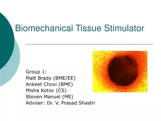
Biomechanical Tissue Stimulator Matthew Brady (BME/EE), Ankeet Choxi (BME), Misha Kotov (CS), Steven Manuel (ME) Vanderbilt University, School of Engineering, Department of Biomedical Engineering, Nashville, TN Advisor: Dr. V. Prasad Shastri. STIMULATION DEVICE. RESULTS AND DISCUSSION.

Biomechanical Tissue Stimulator
E N D
Presentation Transcript
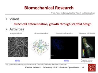
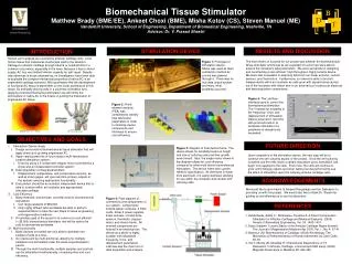
Biomechanical Tissue Stimulator Matthew Brady (BME/EE), Ankeet Choxi (BME), Misha Kotov (CS), Steven Manuel (ME) Vanderbilt University, School of Engineering, Department of Biomedical Engineering, Nashville, TN Advisor: Dr. V. Prasad Shastri STIMULATION DEVICE RESULTS AND DISCUSSION INTRODUCTION Human joint surfaces are covered by articular cartilage (AC), a low friction tissue that transduces mechanical load to the skeleton. Damage to articular cartilage through trauma or osteoarthritis is a common occurrence, especially in the knee. Because it has no blood supply, AC has very limited intrinsic capacity for self-repair. Despite new advances in tissue engineering, no investigators have been able to duplicate the complex mechanical properties of native AC in an engineered cartilage construct. We hypothesize that the development of functional AC tissue is dependent on the zonal architecture of this tissue. By artificially placing cells in a columnar orientation and applying controlled mechanical stimulation we will mimic the architecture of native AC in the hopes of guiding the maturation of engineered AC tissue. The main metric of success for our project was whether the biomechanical tissue stimulator functioned as we expected it to and if we were able to acquire the necessary data parameters. We were successful in designing and constructing a cost-efficient, high-throughput, highly tunable device. We were also successful in acquiring data from our linear encoder, contact sensors, and thermistors. Furthermore, our device is able to function independently within an incubator as cells grow with signals being carried out of the incubator with ribbon wire to an external box housing our electrical and data acquisition components. Figure 1: Prototype of stimulation device. Nitinol was used as main contraction method as current was passed through it. There was no true data output system, and many other problems occurred. Figure 5: The LabView interface used to control the biomechanical stimulator. The VI allows for tunability of the frequency, force, and displacement of stimulation. Data is acquired in real time with protocols built-in to terminate stimulation if a parameter is dangerously exceeded. Figure 2: Finite element analysis (FEA) was completed to identify final fabrication parameters in order to minimize device components and thickness to ensure cost-efficiency. High density AC column AC coverage sites OBJECTIVES AND GOALS FUTURE DIRECTION • Stimulation Device Goals • Design and construct biomechanical tissue stimulator that will apply stimulus to growing engineered AC. • Apply varying amounts of force using a multi-faceted and tunable stimulation system. • Done by using a 3-component stepper motor controlled by a driver and an independent controller system. • Data acquisition of integral parameters • Displacement, temperature, and compression sensors, as well as strain gages, will give real-time primary outputs of the system, ensuring appropriate functionality. • End product should be an isolated, independent device that is able to function within an incubator and appropriately stimulate cartilage. • Cost Efficiency • Many methods, and devices, currently exist for biomechanical stimulation. • Can range upwards of $50,000. • Only highly affluent labs worldwide are able to perform experimentation to take the next steps in tissue engineering and regenerative medicine. • An ancillary goal of the project is to construct a cost efficient (< $5,000) biomechanical stimulator that will be able to be sold to laboratories worldwide. • Multi-functionality • Many devices on market are only able to stimulate one sample of cells at a time. • Our device will be multi-functional, allowing for multiple samples to be stimulated under the same experimentation period. • Through the multi-functionality, multiple samples and controls can be stimulated simultaneously, increasing time and cost efficiency. Figure 3: Diagram of final device frame. The device allows for tunability based on height and size of culturing cells and their growth environment. Also, the single motor shown in the diagram allows for cost efficiency compared to other multi-driven biomechanical stimulators. The device frame was custom milled to specification. As the frame is made from aluminum, it is easily sterilized, allowing for use within the incubator and contact with culturing cells. • Upon completion of the stimulation device, the next step will be to continue the cell-culturing aspect of the project. Once the cell-culturing is started and the cells reach a certain maturation point, stimulation can begin to be applied to the cells. From here, the cells will continue to grow until histology reports and other testing can be performed to see the effect of stimulation upon the culturing articular cartilage cells. ACKNOWLEDGEMENTS We would like to give thanks to Sanjeet Rangarajan and Ian Dalmeyer for providing us with this project. We would also like to thank Dr. Shastri for guiding us and allowing us to use his laboratory. Figure 4: Flow diagram of connections and components of our system. Components include laptop computer, 2 DAQ cards, driver,2 power supplies, linear encoder, contact/force sensors, thermistor, stepper-motor, and device frame. All external components are housed in an electrical box. The device as a whole is highly tunable, with adjustable frequency, force, and displacement parameters. LabView was the main tool of data acquisition and analysis. REFERENCES 1. Aufderheide, Adam C., Athanasiou, Kyriacos A. A Direct Compression Stimulator for Articular Cartilage and Meniscal Explants. (2006) Annals of Biomedical Engineering, Vol. 34. 1463-1474 2. Bobic,Vladimir. Current Status of the Articular Cartilage Repair biomed: The Journal of Regenerative Medicine Apr 2000, Vol. 1, No. 4: 37-41 3. Mansour JM. Biomechanics of Cartilage. (2004) Kinesiology: The Mechanics & Pathomechanics of Human Movement by Carol Oatis. 66-79. 4. Xia Y, Moody JB, Alhadlaq H. Orientational Dependence of T2 Relaxation in Articular Cartilage: a microscopic MRI study. (2002) Magnetic Resonance in Medicine 48: 460-469