Download
1 / 29
290 likes | 528 Views
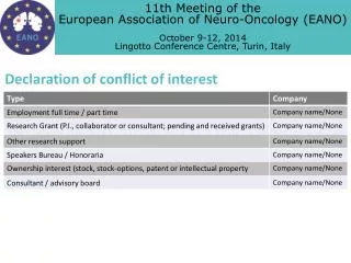
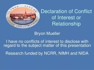
Declaration of Conflict of Interest or Relationship. Speaker Name: Richard G. Spencer I have no conflicts of interest to disclose with regard to the subject matter of this presentation. Unmet Needs for MR in the Unsolved Problem of Tissue Engineering:.

E N D
Declaration of Conflict of Interest or Relationship Speaker Name: Richard G. Spencer I have no conflicts of interest to disclose with regard to the subject matter of this presentation.
Unmet Needs for MR in the Unsolved Problem of Tissue Engineering: Not just one problem, not just one solution Richard G. Spencer Magnetic Resonance Imaging and Spectroscopy Section National Institute on Aging, NIH Baltimore, MD ISMRM Toronto, 2008
Definition of Tissue Engineering: "…an interdisciplinary field that applies the principles of engineering and life sciences towards the development of biological substitutes that restore, maintain, or improve tissue function." Langer and Vacanti, 1993
Tissue Engineering Includes: Implanted cellular constructs designed in the lab --off-the-shelf cartilage, pancreas, liver,… Implanted cells which will develop function in vivo --stem cells for MI or stroke repair --cartilage matrix from implanted cells Implanted acellular scaffolds --bioactive bone graft scaffold Excludes: Organ transplants Artificial mechanical organs Metal joints Hemodialysis, external blood oxygenators,…
Motivation for TE Limitations of non-biologic repair: Duration < longevity of patient (metal implants) Highly invasive (hemodialysis) Typically, no growth or adaptation (c.f. advantage of demand pacemakers)
Funding Opportunities NIH: regenerative medicine funding 2008-- US$575 M (approx. 2% of NIH budget) DoD: Armed Forces Institute of Regenerative Medicine (AFIRM), April, 2008: US$265 M over 5 years Other: NIST, NSF, Arthritis Foundation, Musculoskeletal Transplant Foundation…
The product list is limited but active Medtronic: INFUSE bone grafts RESTORE small intestine submucosa (duPont) Apligraf skin patch (Organogenesis) Cord blood stem cell banking
(SIS) Lysaght, Tissue Engineering, 2008
Private sector activity, 2007 (worldwide) # of firms or business units > 50 # of employees > 3000 Annual sales > US$1.3 billion # of patients treated > 1M # products in preclinical stages > 50 Lysaght, Tissue Engineering, 2008
What Can MRI and MRS Contribute? Non-invasive Monitoring after Implantation Positional stability Integration with surroundings Metabolic activity Need for and response to ongoing intervention "Structural imaging" may be less important than other capabilities of MR, such as more specific microstructural and molecular analyses, and metabolic analysis
R = 0.81, p < 0.001 PCr ATP MDP Pi TR (s) 2.5 31P spectrum of avian neocartilage TE Cartilage Development in Hollow-fiber Bioreactor Bovine neocartilage developing from chondrocytes MT of TE Cartilage 1 week 2 weeks 3 weeks 4 weeks Blue: MT< 0.4 Red: MT> 0.8
Collagen/ Dry Wt. S-GAG/ Dry Wt. Dynamic Modulus Equilibrium Modulus FCD Effect of chondroitinase on developing cartilage Neocartilate in Bioreactor Effect of ibuprofen on developing cartilage Prussian blue stained section MR Image Matrix containing iron-labeled cells Day 14 Non-labeled matrix
Metabolic Mapping in TE cartilage Non MRI: Model Based Estimates (Sengers B. G. et al. JBME 2005) 1H Localized Spectroscopy in Collagen I Constructs Reference geometry of collagen I gel Time course of glucose diffusion into ROI_1 glucose diffusion front culture medium collagen I gel time Computed spatial glucose concentration Fick’s law internal chemical shift standard TSP 10mM 5x5x1 mm voxel Michaelis-Menten Direct measurement of spatial glucose concentration Courtesy David Reiter
Cartilage in hydrogel Biochem vs MRI defect repair tissue native cartilage 10 mM CuCl2 landmark bulb Defect 1 9 days 21 days 28 days 43 days Subchondral bone T2 = 83.4 ms repair tissue defectregion Cartilage/Hydrogel Studies 100µm Cartilage Tidemark Defect 2 Hydrogel defect repair in rabbit Histology vs MRI T2 = 49.5 ms repair tissue defect region 100µm Courtesy S. Ramaswamy, Bioengineering, Univ of Pittsburgh
Asssessment of Cartilage formed in Self-Aggregating Suspension Culture Characteristics of Culture Model Grows in suspension at high density (1-2 X10⁷ cells/ml) Forms a mass quickly (110 weeks mass >6 fold) Maintains cartilage phenotype (cartilage specific ECM, no Coll I ) Bovine patellar cartilage Engineered cartilage Previous studies: Direct correlation of T1ρ MR measurements with FCD and PG in cartilage T1ρ = 107 ms T1ρ = 105 ms MR-based methods offer advantages of providing noninvasive, nondestructive, and quantitative surrogate measures of material properties. Courtesy of George Dodge Bone & Cartilage Research Lab, Nemours Biomedical Research
MRI Following Matrix-assisted Autologous Chondrocyte Transplantation T2-weighted fast spin echo images reveal longitudinal changes in repair zone containing implanted chondrocytes 4 Weeks Post-Surgery: Fluid-like contrast in repair zone 24 Weeks Post-Surgery: Heterogeneous contrast 52 Weeks Post-Surgery: Repair zone isointense with native cartilage Source: Trattnig et al., 2007
Ostogenesis in a Tissue Engineered Construct T1 weighted image of osteochondral plug MSC’s from adult bone marrow gelatin sponge osteogenic cx medium T1 and T2 values for developing TE bone Conclusion: T2 is an effective marker for ossification 11.7 T SE; TE/TR=1000/30 Courtesy Richard Magin, UIC
Non-invasive MRI of tissue-engineered bladder construct Major challenge to successful regeneration: creating immediate angiogenesis Role for imaging: non-invasive quantitative assessment of angiogenesis Angiogenesis at different VEGF levels CD31-immunostaining Exposed bladder construct fat VEGF microvessel density Courtesy of Hai-Ling Margaret Cheng, PhD The Hospital for Sick Children and University of Toronto
Tissue-engineered bladder construct Gadomer (blood pool cx agent): correlation with microvessel density (MVD) through either fractional plasma volume (vp) or area under uptake curve (AUC); superior to Gd-DTPA Gd-DTPA (extracellular cx agent): correlation with MVD is best obtained with AUC SIGNIFICANCE: MRI measurement of absolute blood volume is feasible in therapeutic angiogenesis paradigms Gadomer Gadomer Gd-DTPA Gd-DTPA r = 0.784, P = 0.003 AUC1min vp (%) r = 0.228, P = 0.50 r = 0.437, P = 0.179 r = 0.696, P = 0.012 MVD (%) MVD (%)
MR Monitors Viable Cell Number, Bioenergetics and Oxygen Distribution for Mouse Insulinoma bTC Cells in Tissue-Engineered Pancreas 31P 1D CSI yields bioenergetic data with both spatial and temporal resolution. T1 of 19F PFTBA shows changes in dissolved oxygen content during hypoxic intervention. Constantinidis et al.
Stem Cell Therapeutics—Cell Persistence Loss of cells from noninfarcted tissue Hypointense regions showing sites of mesenchymal stem cell injection D. Kraitchman--JHU
Tissue properties can often be evaluated by highly sensitive but invasive techniques • Gene expression analysis for genotype and tissue quality • Electron microscopy for microstructure • Histology for exquisite structural detail • Immunohistochemistry for protein expression • Optical spectroscopy for more specific matrix components • Microelectrodes / biochemical analysis for metabolic analysis • Mechanical testing for functional support properties
Unmet Needs • Replacement of gold-standard techniques with non-invasive versions • Molecule-specific MR: • example: molecular imaging for collagen I vs collagen II • Mechanical measurements: • example: bone/cartilage elastography; strain measurements • Sequences and analyses more specific to tissue components • example: water compartment analysis; multiparametric approaches • Functional analysis (esp. metabolism) • example: localized spectroscopy for cell number and viability • Histologic-like resolution • example: microscopy
Caveat: Capital value of publicly traded TE companies: 2007: $4.7 billion US 2000: $2.5 billion US 2003: $300 million US Lysaght, Tissue Engineering, 2008
Conclusions Tissue engineering is currently in a growth phase Fundable through several sources There is a substantial market presence (contrast gene therapy) MR can actually contribute to further progress Come to the educational session in Hawaii!