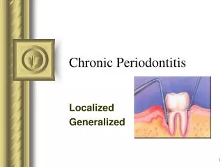
Chronic Periodontitis
This presentation will probably involve audience discussion, which will create action items. Use PowerPoint to keep track of these action items during your presentation In Slide Show, click on the right mouse button Select “Meeting Minder” Select the “Action Items” tab

Chronic Periodontitis
E N D
Presentation Transcript
This presentation will probably involve audience discussion, which will create action items. Use PowerPoint to keep track of these action items during your presentation • In Slide Show, click on the right mouse button • Select “Meeting Minder” • Select the “Action Items” tab • Type in action items as they come up • Click OK to dismiss this box • This will automatically create an Action Item slide at the end of your presentation with your points entered. Chronic Periodontitis Localized Generalized
Learning Outcomes • Describe the development of a periodontal pocket. • Relate clinical characteristics to the histopathologic changes for chronic periodontitis. • Compare the gingival pocket with the periodontal pocket. • Determine the severity of PD activity using clinical data.
Common Characteristics • Onset - any age; most common in adults • Plaque initiates condition • Subgingival calculus common finding • Slow-mod progression; periods of rapid progression possible • Modified by local factors/systemic factors/stress/smoking
Extent & Severity • Extent: • Localized: 30% of sites affected • Generalized > 30% of sites affected • Severity:entire dentition or individual teeth/site • Slight = 1-2 mm CAL • Moderate = 3-4 mm CAL • Severe = 5 mm CAL
Clinical Characteristics • Deep red to bluish-red tissues • Thickened marginal gingiva • Blunted/cratered papilla • Bleeding and/or suppuration • Plaque/calculus deposits
Clinical Characteristics • Variable pocket depths • Horizontal/vertical bone loss • Tooth mobility
Pathogenesis – Pocket Formation • Bacterial challenge initiates initial lesion of gingivitis • With disease progression & change in microorganisms development of periodontitis
Pocket Formation • Cellular & fluid inflammatory exudate degenerates CT • Gingival fibers destroyed • Collagen fibers apical to JE destroyed infiltration of inflammatory cells & edema • Apical migration of junctional epithelium along root • Coronal portion of JE detaches
Pocket Formation • Continued extension of JE requires healthy epithelial cells! • Necrotic JE slows down pocket formation • Pocket base degeneration less severe than lateral
Pocket Formation • Continue inflammation: • Coronal extension of gingival margin • JE migrates apically & separates from root • Lateral pocket wall proliferates & extends into CT • Leukocytes & edema • Infiltrate lining epithelium • Varying degrees of degeneration & necrosis
Continuous Cycle! • Plaque gingival inflammation pocket formation more plaque
Histopathology • Connective Tissue: • Edematous • Dense infiltrate: • Plasma cells (80%) • Lymphocytes, PMNs • Blood vessels proliferate, dilate & are engorged • Varying degrees of degeneration in addition to newly formed capillaries, fibroblasts, collagen fibers in some areas
Histopathology • Periodontal pocket: • Lateral wall shows most severe degeneration • Epithelial proliferation & degeneration • Rete pegs protrude deep within CT • Dense infiltrate of leukocytes & fluid found in rete pegs & epithelium • Degeneration & necrosis of epithelium leads to ulceration of lateral wall, exposure of CT, suppuration
Clinical : Pocket wall bluish-red Smooth, shiny surface Pitting on pressure Histopathology: Vasodilation & vasostagnation Epithelial proliferation, edema Edema & degeneration of epithelium Clinical & Histopathologic Features
Clinical: Pocket wall may be pink & firm Bleeding with probing Pain with instrumentation Histopathology: Fibrotic changes dominate blood flow, degenerated, thin epithelium Ulceration of pocket epithelium Clinical & Histopathologic Features
Clinical : Exudate Flaccid tissues Histopathology: Accumulation of inflammatory products Destruction of gingival fibers Clinical & Histopathologic Features
Root Surface Wall • Periodontal disease affects root surface: • Perpetuates disease • Decay, sensitivity • Complicates treatment • Embedded collagen fibers degenerate cementum exposed to environment • Bacteria penetrate unprotected root
Root Surface Wall • Necrotic areas of cementum form; clinically soft • Act as reservoir for bacteria • Root planing may remove necrotic areas firmer surface
Classification of Pockets • Gingival: • Coronal migration of gingival margin • Periodontal: • Apical migration of epithelial attachment • Suprabony: • Base of pocket coronal to height of alveolar crest • Infrabony: • Base of pocket apical to height of alveolar crest • Characterized by angular bony defects
Periodontal Pocket • Suprabony pocket
Inflammatory Pathway • Stages I-III – inflammation degrades gingival fibers • Spreads via blood vessels: • Interproximal: • Loose CT transseptal fibers marrow spaces of cancellous bone periodontal ligament suprabony pockets & horizontal bone loss transseptal fibers transverse horizontally
Inflammatory Pathway • Interproximal: • Loose CT periodontal ligament bone infrabony pockets & vertical bone loss transseptal fibers transverse in oblique direction
Inflammatory Pathway • Facial & Lingual: • Loose CT along periosteum marrow spaces of cancellous bone supporting bone destroyed first alvoelar bone proper periodontal ligament suprabony pocket & horizontal bone loss
Inflammatory Pathway • Facial& Lingual: • Loose CT periodontal ligament destruction of periodontal ligament fibers infrabony pockets & vertical or angular bone loss
Periodontal Pathogens • Gram negative organisms dominate • P.g., P.i., A.a. may infiltrate: • Intercellular spaces of the epithelium • Between deeper epithelial cells • Basement lamina
Periodontal Pathogens • Pathogens include: • Nonmotile rods: • Facultative: • A.a., E.c. • Anaerobic: • P. g., P. i., B.f., F.n. • Motile rods: • Facultative: • C.r. • Spirochetes: • Anaerobic, motile: • Treponema denticola
Periodontal Disease Activity • Bursts of activity followed by periodsof quiescence characterized by: • Reduced inflammatory response • Little to no bone loss & CT loss • Accumulation of Gram negative organisms leads to: • Bone & attachment loss • Bleeding, exudate • May last days, weeks, months
Periodontal Disease Activity • Period of activity followed by period of remission: • Accumulation of Gram positive bacteria • Condition somewhat stabilized • Periodontal destruction is site specific • PD affects few teeth at one time, or some surfaces of given teeth
Overall Prognosis • Dependent on: • Client compliance • Systemic involvement • Severity of condition • # of remaining teeth
Prognosis of Individual Teeth • Dependent on: • Attachment levels, bone height • Status of adjacent teeth • Type of pockets: suprabony, infrabony • Furcation involvement • Root resorption