Download
1 / 1
10 likes | 132 Views
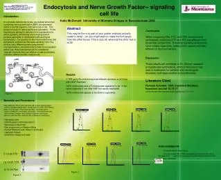
This project explores the role of signaling endosomes in the process of endocytosis and their effect on nerve growth factor (NGF) signaling within cells. We analyze how specific endosomes are formed, focusing on whether they possess unique characteristics that differentiate them from one another, particularly in size, density, and protein composition. Results indicate that endosomes containing different proteins like P75 and pTrk exhibit distinct characteristics, contributing to ongoing research into treatments for neurological diseases.

E N D
Endocytosis and Nerve Growth Factor-- signaling cell life The University of Montana Division of Biological Sciences Introduction In a process called endocytosis, specialized edosomes containing nerve qrowth factor (NGF) are generated within a cell that signal for that cell to live or die. This project focuses on data analysis and generation. These experiments attempt to determine the mechanisms by which signaling endosomes are formed and what proteins participate in membrane traffic and signal transduction. Our question is: Are there endosomes that are special, endosomes that keep signaling? And if so, do they carry different protein components? Our hypothesis is, we believe the answer to our question will be yes, these endosomes will be considered “special”, because they are different in size and density and carry different protein components. Katie McDonald University of Montana Bridges to Baccalaureate 2006 Abstract This may be the only part of your poster anybody actually reads in detail – so you might want to make the font larger than the other boxes. This is size 36, whereas the other text is at 32. Conclusion When comparing Ptrk, P75, and TFR, we found that endosomes containing P75 and TFR are different from those that contain Ptrk. Therefore signaling endosomes have unique organelles, making them special and also different in size and density. Discussion These results will contribute to Dr. Grimes’ research proposals and publications, which in the future may lead to treatments for patients with neurological diseases, both degenerative and proliferative. Results • TFR and pTrk endosomes had different densities at all times after NGF treatment • pTrk endosomes and p75 endosomes appeared to be in the same organelle 2 min after NGF but rapidly separated • pTrk endosomes appear to be distinct organelles Literature Cited Famous Scientist. 1999. Important literature. Important Journal 12:13-17. Famous Scientist. 1999. Important literature. Important Journal 12:13-17. Figure 1. • Materials and Procedures • Initial Materials: PC12 cells (rat brain cell), pipets, blotting paper, • dd H2O (double distilled H2O), normal wash (1x TBS .05% Tween), • Blotto (normal wash + 5% nonfat dry milk), secondary wash • (blotto + antibiotin + secondary rat/mouse), luminal and peroxide, • plastic dishes, shakers, glass plates, incubator, centrifuges, • dark box, and the iMac computer with software for quantifying. • Harvested cells • Centrifugation (cell fractionation) • Generated endyzomes • Ran endyzomes on Western Blots • Probed Westerns with different antibodies • Captured images • Quantified images and data Acknowledgmnts Research Mentor: Mark Grimes Funding: Project IBS-CORE Undergraduate Research Fellowship, provided by a grant from the Howard Hughes Medical Institute to the University of Montana. 5-10-06 P75 12-13-05 TFR 6-13-05 Ptrk Figure 3. Figure 2.