Download
1 / 18
210 likes | 733 Views
Embryonic and Fetal Development of Respiratory System. Fred Hill, MA, RRT. Where Do Babies Come From?. "There are only two ways to live your life. One is as though nothing is a miracle. The other is as though everything is a miracle." Albert Einstein. Duration of Human Preganancy.

E N D
Embryonic and Fetal Development of Respiratory System Fred Hill, MA, RRT
Where Do Babies Come From? • "There are only two ways to live your life. One is as though nothing is a miracle. The other is as though everything is a miracle." • Albert Einstein
Duration of Human Preganancy • 10 lunar months • 9 calendar months • Three trimesters (3 months each) • 40 weeks
Stages of Growth and Development • Conception to completion of implantation (12 to 14 days) • Ovum • Embryonic Development (to 54-56 days) • Embryo • Vulnerable • Major organ systems are developed • Fetal Stage (to end of pregnancy) • Birth • Neonate (to one month) • Infant (to one year) • Child (>1 year)
Germ Layers and Associated Organs • Ectoderm (outer layer) • Epidermis • Hair, nails • Lens of eye • Nervous system • Skin glands • Endoderm (inner layer) • Respiratory tract • Epithelium of digestive tract, bladder, thyroid • Liver and pancreas • Mesoderm (middle layer) • Dermis • Muscles • Bone, connective tissue. lymphoid tissue • Reproductive organs • Cardiovascular system
Lung Development • Embryonic period • 24 days: lung bud appears • 28 days: right and left lung buds • 31 days: lobar branches appear • Diaphragm formed by 7 weeks
Pseudoglandular period (7–16 weeks) • 7 weeks • choanae form between nasal cavity and oropharynx • Separation of oral and nasal cavities begins • 8 weeks: vocal cords appear • 12 weeks: palates completely formed • Majority of airway branching occurs weeks 10-14 • 13 weeks • goblet cells are formed • Bronchial gland formation begins
Canalicular period (17-26 weeks) • Terminal and respiratory bronchioles continue to develop • Airways increase in length and diameter • Few pulmonary capillaries present early in period • Pulmonary capillaries proliferate toward end of period • Formation of alveolar ducts • Appearance of alveolar type I and II cells • 24-26 weeks: capillaries close enough to allow gas exchange • 24 weeks: bronchial glands complete development • According to a brief submitted to the Supreme Court in WEBSTER V. REPRODUCTIVE HEALTH SERVICES by more than 150 distinguished scientists and physicians, "There are no medical developments anticipated in the foreseeable future that would bring about adequate fetal lung function prior to 23 or 24 weeks of gestation."
Sacular and alveolar period (27-40 weeks) • 32-34 weeks: True alveoli appear • Number of alveoli continue to increase until about age 8 years • 35 weeks: mature surfactant is present. PG is present in surfactant.
Surfactant • Appearance coincides with development of Type II cells (week 17) • Lowers surface tension to decrease muscular effort to open and ventilate the lungs • Mature surfactant noted by: • Presence of phosphatidylglycerol (PG) • L/S ratio > 2:1
Lung Profile • Presence of PG • L/S ratio • Conditions that delay or accelerate surfactant production (see Table 1-2) • Lack of surfactant is leading cause of pulmonary complications in the newborn
Fetal Lung Fluid • At term, fetal lung holds ~ 20-30 ml/kg • Increasing levels of fluid production in late gestation – exits lung and excreted through mouth into amniotic fluid • Different composition than amniotic fluid: ↓ pH, ↓ protein, ↓ HCO3, ↑ Na+, ↑ Cl-
Fetal Circulation • Fetal shunts • Ductusarteriosus • Foramen ovale • Ductusvenosus • Placenta – gas exchange, not lungs • Right heart pressures > left • High PVR • Low SVR • Umbillical vessels • Arteries: carry deoxygenated blood • Vein: carries oxygenated blood
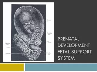
Intrauterine Structures • Placenta: Gas exchange & waste removal for developing fetus • Umbilical cord • Three vessels: two arteries & one vein • Wharton’s jelly • Amnion: membrane containing fetus and amniotic fluid • Oligohydramnios • Polyhydramnios
Amniotic Fluid • Amniotic fluid is constantly being produced and absorbed • Past 24-26 weeks the fetus swallows amniotic fluid • Produced by fetal urination and lung secretion • Functions • Allows fetal movement and growth • Protects from traumatic injury • Thermoregulation/homeostasis