Download
1 / 19
200 likes | 449 Views
D-Influenza virus. Influenza epidemiology in humans. Fields Virology, 2nd ed, Fields & Knipe, eds, Raven Press, 1990, Fig.40-1. Influenza mortality from 1957 to 1979. Fields Virology, 2nd ed, Fields & Knipe, eds, Raven Press, 1990, Fig.40-11. Influenza virus structure.

E N D
Influenza epidemiology in humans Fields Virology, 2nd ed, Fields & Knipe, eds, Raven Press, 1990, Fig.40-1
Influenza mortality from 1957 to 1979 Fields Virology, 2nd ed, Fields & Knipe, eds, Raven Press, 1990, Fig.40-11
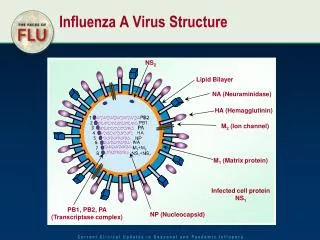
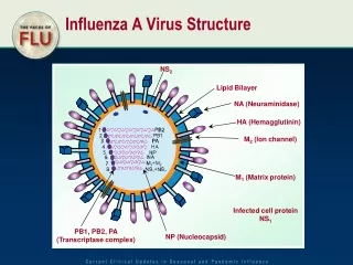
Influenza virus structure Structure of influenza virus. The diagram illustrates the main structural features of the virion. The surface of the particle contains three kinds of spike proteins: the hemagglutinin (HA), neuraminidase (NA), and matrix (M2) protein embedded in a lipid bilayer derived from the host cell and covers the matrix (M1) protein that surrounds the viral core. The ribonucleoprotein complex making up the core consists of at least one of each of the eight single-stranded RNA segments associated with the nucleoprotein (NP) and the three polymerase proteins (PB2, PB1, PA). RNA segments have base pairing between their 3´ and 5´ ends forming a panhandle. Their organization and the role of NS2 in the virion remain unresolved. (From Fields Virology, 4th ed, Knipe & Howley, eds, Lippincott Williams & Wilkins, 2001, Fig. 47-2)
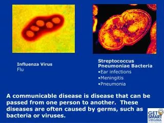
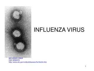
Influenza virus Electron micrographs of influenza virus. A–C: The structure of the internal components; (D) the external view. A substantial fraction (up to 50%) of influenza virions contain large helical internal components (A, B), which may contain individual ribonucleoprotein (RNP) segments (C) linked together. The individual RNPs each contain a binding site for the viral polymerase, as seen by the immunogold labeling of the end of the RNP segment (C). The external view of the virions (D) illustrates the pleomorphic appearance and the surface spikes. Bar in all figures equals 50 nm. (From Fields Virology, 4th ed, Knipe & Howley, eds, Lippincott Williams & Wilkins, 2001, Fig. 47-2)
Influenza gene functions From Medical Microbiology, 5th ed., Murray, Rosenthal & Pfaller, Mosby Inc., 2005, Table 60-1.
Structure of the influenza hemagglutinin monomer HA monomer. Sites A-E are immunodominant epitopes (From Fields Virology, 2nd ed, Fields & Knipe, eds, Raven Press, 1990, Fig.40-4)
Structure of the influenza hemagglutinin trimer HA trimer. (From Fields Virology, 2nd ed, Fields & Knipe, eds, Raven Press, 1990, Fig.39-6)
Influenza A hemagglutinin and neuraminidase subtypes Fields Virology, 4th ed, Knipe & Howley, eds, Lippincott Williams & Wilkins, 2001, Table 47-1
Influenza A reservoir The reservoir of influenza A viruses. The working hypothesis is that wild aquatic birds are the primordial reservoir of all influenza viruses for avian and mammalian species. Transmission of influenza has been demonstrated between pigs and humans (solid lines). There is extensive evidence for transmission between wild ducks and other species, and the five different host groups are based on phylogenetic analysis of the nucleoproteins of a large number of different influenza viruses. (From Fields Virology, 4th ed, Knipe & Howley, eds, Lippincott Williams & Wilkins, 2001, Fig. 47-3.)
Influenza replication From Medical Microbiology, 5th ed., Murray, Rosenthal & Pfaller, Mosby Inc., 2005, Figure 60-2.
Influenza pathogenesis in humans Six seronegative volunteers received 104.0 TCID50 of wild-type A/Bethesda/1015/68 virus intranasally on day 0. (From Fields Virology, 4th ed, Knipe & Howley, eds, Lippincott Williams & Wilkins, 2001, Fig. 47-10.)
Influenza pathogenesis From Medical Microbiology, 5th ed., Murray, Rosenthal & Pfaller, Mosby Inc., 2005, Figure 60-3.
Influenza reassortment in an intermediate host Natural demonstration of reassortment of influenza variants in an intermediate host, with subsequent interspecies transmission. A 1980 H7N7 emergent virus with high mortality for seals is consistent with reassortment between two concurrently circulating avian viruses. The virus caused a conjunctivitis in humans handling the seals, and could be experimentally transmitted (assymptomatically) to other species. (From Fields Virology, 2nd ed, Fields & Knipe, eds, Raven Press, 1990, Fig.40-8)
Influenza reassortment in humans Genetic reassortment between human H1N1 and H3N3 viruses. Reassortamt viruses (R1 and R2) containing mixed gene constellations were isolated from humans during 1978 and 1979 but not since. (From Fields Virology, 2nd ed, Fields & Knipe, eds, Raven Press, 1990, Fig.40-9)
Antigenic drift Antigenic drift of H3N2 viruses between 1968 and 1997 as demonstrated by their cross-reactivity in hemagglutination-inhibition tests. (From Fields Virology, 4th ed, Fields & Knipe, eds, Raven Press, 1990,Table 47-3.)
Influenza A evolution: antigenic shift Postulated evolution of the influenza A viruses currently circulating in humans. Seroarcheology suggests that H2N2 and H3N8 influenza viruses circulated in humans in 1889 and 1900, respectively. Phylogenetic evidence suggests that an influenza virus possessing eight gene segments from avian influenza reservoirs was transmitted to humans and pigs before 1918 and replaced the 1900 strain. This virus was probably carried from North America to Europe by American troops and caused the catastrophic Spanish influenza pandemic of 1918. In 1957 the Asian pandemic virus acquired three genes (PB1, HA, and NA) from the avian influenza gene pool in wild ducks by genetic reassortment and kept five other genes from the circulating human strain. After the Asian strain appeared, the H1N1 strains disappeared from humans. In 1968 the Hong Kong pandemic virus acquired two genes (PB1 and HA) from the duck reservoir by reassortment and kept six genes from the virus circulating in humans. After the appearance of the Hong Kong strain, the H2N2 Asian strains were no longer detectable in humans. In 1977 the Russian H1N1 influenza virus that had circulated in humans in 1950 reappeared and spread in children and young adults. This virus probably escaped from a laboratory and has continued to cocirculate with the H3N2 influenza viruses in the human population. (From Fields Virology, 4th ed, Knipe & Howley, eds, Lippincott Williams & Wilkins, 2001, Fig. 47-1.)
Summary: influenza • Structure • Negative sense segmented ssRNA genome, helical nucleocapsid, enveloped • Pathogenesis • respiratory transmission • replication in nucleus; budding • no spread (usually) • innate and antibody response important; antigenic shift and drift • local symptoms from cell killing; systemic symptoms from immune response; exaggerated disease in young and elderly; viral and bacterial pneumonia complications • Diagnosis • culture, hemadsorbtion, viral antigen detection • Treatment/prevention • amantidine and rimantidine target matrix; zanamivir and oseltamivir target NA • killed and live vaccines need constant updating