Download
1 / 39
460 likes | 675 Views
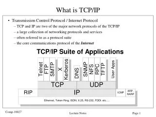
PET/CT Protocol, Image Reconstruction and Data Transfer for Radiotherapy. DR. Halima Hawesa RAD 466 Lecture 6. Presentation Out Lines. PET/CT System PET/CT System Design PET/CT Instrumentations PET/CT Imaging Method New Advances in PET PET/CT Imaging protocols

E N D
PET/CT Protocol, Image Reconstruction and Data Transfer for Radiotherapy DR. Halima Hawesa RAD 466 Lecture 6
Presentation Out Lines • PET/CT System • PET/CT System Design • PET/CT Instrumentations • PET/CT Imaging Method • New Advances in PET • PET/CT Imaging protocols • PET/CT Processing Parameters • CT Acquisition recommendations • PET/CT Acquisition recommendations • Contrast medium injection • PET/CT for external RT planning • PET/CT for therapy monitoring • Alternative PET tracer imaging • Possible PET/CT Pitfalls • Data transfer, data format and data security • Combined image display in PET/CT • Clinical applications of PET/CT • Present and future of PET/CT system
PET System Characteristics • Positron Emission Tomography (PET), as an integral part of nuclear medicine imaging, is a non-invasive imaging technique offering the visualization and quantification of pre-selected tracer metabolism • All commercial PET systems today come as a full ring arrangement of detector blocks or curved pixel arrays, both covering an axial imaging range of up to 23 cm. • The scintillators-of-choice are LSO, or LYSO, and GSO, offering a higher light output and shorter decay times
CT System Characteristics • Computed tomography (CT) is the science that creates two-dimensional cross-sectional images from three-dimensional body structures • The primary goal of any CT system is to accurately reproduce the internal structures of the body as two-dimensional cross-sectional images. • This goal is accomplished by computed tomography's superior ability to demonstrate slight differences in tissue contrast. • Each component of a CT system plays a major role in the accurate formation of each CT image produced
PET AND CT PROBLEMS • The goal of CT imaging is to generate an attenuation map. • Attenuation of the annihilation photons is a major source of error in PET imaging, if uncorrected. • In PET, the matter in the body non-uniformly attenuates photons resulting in incorrect information about the actual quantity of radiotracer. • Without corrections for this physical effect, PET images have quantitatively inaccurate activity concentrations.
PET/CT System Characteristicscross-modality imaging systems • (PET/CT) gains more and more importance as a part of the treatment-planning procedure in radiotherapy. • Imaging technique, offers the visualization and quantification of pre-selected tracer metabolism. • In combination with the structural information from CT, this molecular imaging technique has great potential to support and improve the outcome of the treatment-planning procedure prior to radiotherapy. • By the choice of the PET-Tracer, a variety of different metabolic processes can be visualized. • Glucose metabolism of a tissue is the most metabolite traced by PET, as well as hypoxia or cell proliferation for example.
PET/CT System • PET/CT system is a superior of PET or CT alone only when PET/CT image artifacts are minimized, and accurate quantification of CT and PET results following CT-based attenuation correction is ensured. • All steps of the PET/CT-imaging have to be monitored and the required adjustments have to be considered when ever needed.
PET/CT-Imaging Procedure • Patient preparation • Patient positioning • Scout scan • CT acquisition • PET acquisition • Data processing and reconstruction • Image analysis and reporting
PET/CT Scanner design • Illustration of the major PET/CT scanner components.
PET (A) and PET/CT (B) system design and imaging protocol. (A) PET transmission and emission scanning are performed consecutively and stepwise for a pre-defined number of bed positions. • (B) In combined PET/CT imaging the standard PET transmission scan is replaced by a single, continuous spiral CT scan and the total examination time is shortened to about 20 min. The CT images are used for AC of the emission data.
PET/CT Instrumentation • Today about 75% of all PET systems installed in Europe are PET/CT. • All PET/CT systems for clinical use combine a whole-body PET and a standard, clinical multi-slice CT within a single gantry • Despite early efforts on a fully integrated emission-CT system no fully integrated, single-detector PET/CT exists today • The centers of the fields-of-view of the CT and the PET are axially displaced by up to 85 cm. • Combined PET/CT offers the best possible intra-patient co-registration of complementary anatomical and functional images.
PET/CT adaptations to radiation oncology applications. (A) RT laser bridge installation in front of the combined gantry, (B) patient support system (C) as in (B) with additional thermoplastic head restraint, vacuum lock bag and breast board (from left to right) and (D) dedicated, big-bore PET/CT with an 85-cm gantry opening (Philips Healthcare Systems)
PET/CT System Procedure • patients scheduled for RTP can be positioned on a flat radiation therapy pallet that is mounted easily on the top of the PET/CT patient pallet • A dedicated RT laser system can be installed in front of the PET/CT gantry to ensure proper positioning of the patient based on the pre-defined markers (follow-up scans), or to define the iso-center if the patient is imaged pre-therapy on the PET/CT • With an increased bore diameter adapted to more versatile radiation therapy treatment planning to ensure adequate and reproducible patient positioning using all standard RT-positioning aids during the diagnostic, pre-treatment PET/CT examination
Recent advances in PET technology and impact on PET/CT-guided patient management in radiation oncology.
PET/CT Acquisition and Processing ParametersStandard PET/CT imaging protocol • Most PET/CT imaging protocols today involve so-called wholebody FDG-PET and CT examinations covering the same co-axial imaging range • The use of intravenous contrast medium in PET/CT optimizes the patient’s imaging work-up, characterizes lesions and highlights lesions in some situations. • Some findings such as pulmonary embolism (blockage of the main artery of the lung )will be discovered incidentally in approximately 4% of oncologic patients and probably missed if the examinations are not systematically reviewed by radiologists • If a contrast-enhanced full dose CT is performed it should always be read by a radiologist or CT-trained nuclear medicine physician or a radiation oncologist. • The CT part of the PET/CT, especially if carried out as diagnostic CT (high mAs, contrast media injection), requires specific skills in radiology
PET/CT Acquisition Procedure(Table caption) • (a)Fasting prior the examination is not essential. It is recommended if intravenous contrast medium is given. • (b) Glass of gastrografin or water just before CT acquisition if esophageal or upper GI (Gastrointestinal) tumor. • (c) Two injections are proposed: first for tissue impregnation (To fill throughout), second for vessel. Pacification (elimination). • (d) N refers to the number of detectors: N = 16 if a 16-slice CT is employed. • (f) mAs: absolute values of mAs cannot be recommended in view of significant differences in operating characteristics between CT scanners. The values are given as indicators
PET/CT Acquisition Procedure(Table caption) • (a) Some times it is necessary to follow the tracer kinetics in the brain. Thus, a dynamic protocol must be followed. • The PET-acquisition protocol then consists of several time (f.i. 3 x 5 min).
PET/CT Processing Parameters • The software shipped with modern PET/CT systems often includes several reconstruction algorithms and each of them involves many parameters and options, such as the data corrections, the reconstruction mode (2D, 3D to 2D rebinning, 3D), time-of-flight reconstruction or classical sinogram-based reconstruction . • Data corrections for decay, scatter, attenuation, dead time, depth of interaction. • Reconstruction algorithms can be either analytic or iterative.
PET/CT Processing Parameters • Modern iterative reconstruction algorithms are less prone to artifacts than analytic reconstruction • The main parameters of reconstruction algorithms are the matrix size (or equivalently the voxel size) and the regularization scheme. • Regularization scheme controls the signal-to-noise ratio of the images and encompasses the filter settings in analytic reconstruction.
PET/CT Processing Parameters • The reconstruction algorithm can influence the resolution of the images. For instance, heavy smoothing blurs the images. • The choice of a reconstruction algorithm and the adjustment of its parameters lead to a tradeoff that can influence the presence of artifacts, the signal-to-noise ratio, the resolution, the quantification accuracy, and the reconstruction time. • Depending on the purpose of the images, it can be useful to optimize the reconstruction protocol • High resolution makes partial volume effect easier to correct.
PET/CT Reconstruction Parameters • The outcome of all these applications can be jeopardized by inappropriate or insufficiently optimized reconstruction settings. • In any case, the best reconstruction protocol is of little help if the acquired data are of low quality. • The signal-to-noise ratio of the image is indeed strongly conditioned by the acquisition duration, the tracer specificity, and the injected dose. • Motion blur can be addressed by performing gated acquisitions in modern PET/CT systems
CT-Imaging Parameters • Several CT imaging parameters have to be selected for CT acquisition: slice thickness, tube current and exposure time (mAs), tube voltage (kV), rotation time, pitch, field-of view, reconstruction filter, and image matrix. • Normally, these combinations of parameters are preconfigured and comprised in protocols that can be selected by the operator. • Recent PET/CT systems are equipped with multi-slice CT technology which can acquire up to 128 slices in a single rotation. • This faster scanning speed enables to get more and thinner slices than single- slice CT. In multi-slice CT, almost all different slice thicknesses can be obtained in routine by combining neighboring detectors, ranging from 0.5 to 10 mm. • The mAs directly relates to the number of photons emitted in a X-ray beam, and therefore inversely to noise. • Increasing mAs will decrease noise and increase in contrast resolution with higher dose to the patient and higher heat load on the tube anode.
CT-Imaging Parameters • It has to be noted that doses delivered to the patient can be drastically reduced with a preserved image quality by using anatomical on-line tube dose modulation resulting in an overall decrease in average tube current and in a lower mAs product per rotation. • The kV reflects the energy spectrum of the photons the X-ray beam consists of. Beams of higher photon energy offer superior penetration through the patient so that a larger number of photons reach the detectors. • Tube voltage is usually fixed at 120 kV for general applications. • The rotation time is the time needed for the X-ray beam to complete one 360 revolution. • The benefit of a longer revolution time is the higher mAs it can deliver, leading to increased contrast resolution. However, longer revolution times may also allow more patient motion and, thereby, leads to net loss in image quality. • Rotation time is usually around 0.5–1 s for general purposes. The pitch in spiral scanning is the ratio of the patient’s movement through the gantry during one 360 beam rotation relative to the beam collimation. • The pitch can be defined as follows: pitch = patient (table) advancement per 360 gantry rotation/beam. For pitch > 1, the dose will be reduced. • During the image reconstruction process, the CT data will be passed through mathematical filter algorithms, each optimized for different body parts and tissue types. • For example, a soft tissue algorithm may lead to smooth images while a bone algorithm will sharpen bone margins. • The field-of-view is defined as the maximal diameter of the reconstructed image.
Contrast Medium Injection • The use of intravenous contrast media during CT examination is essential to analyse solid organs, assess vessels and define tumor invasion for treatment planning and organ definition. • The presence of contrast materials on CT images can cause problems in the attenuation correction of PET images. • However, newer research has found that contrast-enhanced CT scans can be used for attenuation correction in oncologic patients with minimal effect in the image quality.
Contrast Medium Injection • Some investigators have proposed CT correction methods to markedly reduce these artefacts. • All intravenous contrast agents currently approved for CT contain iodine bound in a complex organic molecule. • Ionic agents are now abandoned in favour of non-ionic agents. • The contrast medium is injected preferably with a power injector through a flexible catheter placed in the forearm
PET/CT for External RT Planning • Patients should be positioned for the PET/CT in the radiation treatment position when patients will receive external radiation therapy to define the active tumor and target volumes. • External positioning RT lasers can help define the isocentre on PET/CT, which is referenced on the patient and transferred to the treatment plan. • PET/CT-guided treatment planning for lung cancer remains a challenge where respiration- induced misalignments between CT and PET data within a single PET/CT scan can lead to a significant underestimation of the standardized uptake value of the attenuation-corrected PET. • Lung cancer patients require special adaptations to the PET/CT acquisition and treatment delivery. • If external treatment is delivered during normal respiration then adequate verification of the PET/CT data appears sufficient to ensure an accurate representation of the target volume. In case of minimal PET–CT misalignment the mis-registration can be corrected prior to a second attenuation correction using a linear or non-linear co-registration algorithm. • Alternatively, the transmission data could be corrected manually to match the uncorrected emission data, and the CT-AC (CT-based attenuation correction of PET data) could be re-performed.
PET/CT for Therapy Monitoring • In addition to the above-mentioned patient set-up and imaging considerations attention must be given to maintain similar preimaging conditions (blood glucose level, tracer distribution time, and uptake conditions). • By hold on to standard set-up and imaging protocols, reproducible quantitative accuracy of PET/CT can be maintained and accurate therapy response assessment is feasible.
Alternative PET tracer imaging (hypoxia, thyroid and internal dosimetry) • Tracers other than FDG are being increasingly used in defining the active tumor volume. • Examples for these tracers include, but are not limited to, 11C-choline (prostate cancer), 64Cu-ATSM, 18F-FMISO and 18F-FAZA (hypoxia imaging) and 124I for thyroid cancer. • While most tracers are labeled with radioisotopes that have a similar beta fraction as 18F (FDG) some nuclides (124I) create a challenge to the PET detection system and may have implications for calculating a dosimetry plan for internal radiation therapy planning . • PET quantification of impure positron emitters is the work in progress in several research and clinical institutions. PET/CT is ready to be implemented in routine radiation oncology management
Possible PET/CT Pitfalls • Typical pitfalls in standard PET/CT imaging include: - motion-induced mis-registration. -overestimation of PET activity following CT contrast administration. -PET image artifacts in the vicinity of metal implants. • The table in the next slice lists the sources of these artifacts, key studies and suggested solutions.
Sources of Artifacts in Standard PET/CT Imaging -consequences and solutions-
Data Transfer, Data Format and Data Security • Today, radiation-treatment planning (RTP) is based mostly on anatomical image data . Over the past decade, however, treatment plans have started to incorporate also functional image information, which represents various metabolic pathways. • Positron emission tomography (PET) and, more recently PET/CT, based on 18F-FDG were shown to have a significant clinical impact on the diagnosis of a variety of cancers. While 18F-FDG reveals increased glucose metabolism, other compounds can be used for more specific information. • The goal of functional imaging for radiation treatment planning is to add complementary information to anatomical imaging as anatomical imaging is known to be inadequate in certain imaging situations, such as in the differentiation of necrotic from active tumor volume. • PET and PET/CT can serve as a source for explanation of metabolically active target volumes or nasty tumor volume.
Data flow chart in PET/CT system with the hospital PACS system
Combined Image Display in PET/CT • The combination of anatomic and functional images is often called ‘‘fusion.’’ It should be stressed that the images are never ‘‘fused’’ together. • The term ‘‘fusion’’ in this field often refers to the visualization of two distinct images on the same display and is probably better denoted as ‘merged.’’ • There are several methods for merging images on a single display and a common PET/CT method is ‘‘alpha’’ blending. • In this method, the color display values in the PET image are averaged with those of the CT image and the contribution of the PET versus the CT in the final image can be varies. • Side-by-side viewing of the PET emission and CT images with ‘‘locked cursors’’ is a useful technique because overlaying images causes a reduction in the CT contrast
Combined Image Display in PET/CT • Flow chart of a typical PET/CT operation. Attenuation correction factors can be obtained from either (A) CT-based or (B) conventional PET transmission source.
Clinical Applications of PET/CT • Incidental Unsuspected Malignancies • Head and Neck • Brain • Unknown Primary Tumors • Initial Diagnosis • Prognosis • Thyroid Carcinoma • Lung Cancer • Esophageal Cancer • Breast Cancer • Colorectal Cancer • Non-Hodgkin and Hodgkin Lymphoma • Melanoma
Present and Future Summary • PET/CT technology has become extremely popular in the last 4 years, with approximately 400 installed scanners worldwide. • PET/CT offers a simple hardware solution for viewing anatomic and functional images and a significant reduction in emission imaging time because of the use of CT-based attenuation correction. • These same benefits also pose technical challenges. Specifically, the final image alignment is still prone to minor error and the translation of CT values to PET for attenuation correction suffers from several complications. • The future of PET/CT awaits the results from clinical studies evaluating this technology for tasks including diagnosis, serial follow-up, and radiation treatment planning. • In the meantime, PET/CT systems continue to evolve to include improved software and hardware features. New techniques are being explored for CT-guided PET image reconstruction ultimately to improve detection or quantitation.
Reference for this lecture is the pdf file (petct full procedure) Thanks for listening