
Introduction
No. 040. Assessment of p roliferative index and its association with KI -67 antigen molecule expression in benign prostatic hyperplasia. David Low , Srikumar Chakravarthi , Thanikachalam P, Nagaraja HS, Nadeem Irfan Bukhari . International University Malaysia, Kuala Lumpur.

Introduction
E N D
Presentation Transcript
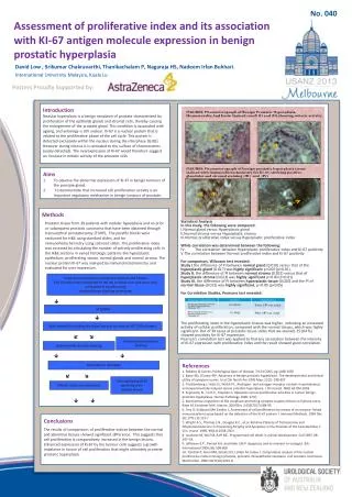
No. 040 Assessment of proliferative index and its association with KI-67 antigen molecule expression in benign prostatic hyperplasia David Low , SrikumarChakravarthi, ThanikachalamP, NagarajaHS, NadeemIrfanBukhari. International University Malaysia, Kuala Lumpur Posters Proudly Supported by: Introduction Nodular hyperplasia is a benign neoplasm of prostate characterized by proliferation of the epithelial glands and stromal cells, thereby causing the enlargement of the prostate gland. This condition is associated with ageing, and aetiologyis still unclear. Ki-67 is a nuclear protein that is related to the proliferative phase of the cell cycle. This protein is detected exclusively within the nucleus during the interphase (G (0)). However during mitosis it is relocated to the surface of chromosomes (easily detected). The overexpression of Ki-67 would therefore suggest an increase in mitotic activity of the prostatic cells Statistical Analysis In this study, the following were compared: Normal gland versus Hyperplastic gland Normal stroma versus Hyperplastic stroma Normal proliferative index versus Hyperplastic proliferative index While correlation was determined between the following: IV. The correlation between Hyperplastic proliferative index and Ki-67 positivity The correlation between Normal proliferative index and Ki-67 positivity For comparison, Wilcoxon test revealed: Study I the difference of PI between normal gland (0.010) versus that of the hyperplastic gland (0.017) was highly significant p=0.00 (p<0.01). Study II, the difference of PI between normal stroma(0.012) versus that of hyperplastic stroma(0.023) was highly significant p=0.00 (P<0.01) Study III, the difference of PI between hyperplastic tissue (0.020) and the PI of normal tissue (0.013) was highly significant, p=0.00 (p<0.05) For Correlation Studies, Pearsons test revealed: The proliferating index in the hyperplastic tissues was higher, indicating an increased activity of cellular proliferation, compared with the normal tissues, which was highly significant. Out of 39 cases of prostatic tissue slides that we stained, 25 (64 %) showed positivity for Ki-67 expression. Pearson’s correlation test was applied to find any association between the intensity of Ki-67 expression with proliferative index and the result showed good correlation. Aims To observe the abnormal expression of Ki-67 in benign tumours of the prostate gland. To demonstrate that increased cell proliferation activity is an important regulatory mechanism in benign tumours of prostate. • Methods • Prostatic tissue from 39 patients with nodular hyperplasia and no prior or subsequent prostatic carcinoma that have been obtained through transurethral prostatectomy (TURP). The paraffin blocks were sectioned for H&E using standard slides, and for immunohistochemistry using salinized slides. The proliferative index was assessed by calculating the number of actively proliferating cells in the H&E sections in varied histologic patterns like hyperplastic epithelium, proliferating stoma, normal glands and normal stroma. The nuclear protein Ki-67 was analyzed by immunohistochemistry and evaluated for over expression. • • • • • • Chips of tissue samples (1cm3) were washed and fixed in 10% formalin and transported to the lab in plastic vials and were then embedded in paraffin using standard tissue blocking techniques. STAINING Wax removal by putting the slides into hot air oven at 56°C (30 minutes) Immunohistochemistry Staining Haematoxylin & Eosin Staining References 1. Robbins & Cotran; Pathological basis of disease; 7th Ed 2005; pg 1048-1050 2. Barry MJ, O'Leary MP: Advances in benign prostatic hyperplasia. The developmental and clinical utility of symptom scores. UrolClin North Am 1995 May; 22(2): 299-307 3. Trachtenberg J, Hicks LL, Walsh PC. Androgen- and estrogen-receptor content in spontaneous and experimentally induced canine prostatic hyperplasia. J Clin Invest. l980; 65:l05l-1059 4. Kyprianou N., Tu H.C., Stephen C. Apoptotic versus proliferative activities in human benign prostatic hyperplasia. Human Pathology 1996; 27(7). 5. Autonomous regulation of the anaphase-promoting complex couples mitosis to S-phase entry. Rape M, Kirschner MW. Nature. 2004 Dec 2;432(7017):588-95. 6. Key G, Kubbutat MH Gerdes J, Assessment of cell proliferation by means of an enzyme- linked immunosorbent assay based on the detection of the Ki-67 protein. J Immunol Methods. 1994 Dec 28; 177(1-2):113-7 7. Wright A.S., Thomas L.N., Douglas R.C., et.al. Relative Potency of Testosterone and Dihydrotestosterone in Preventing Atrophy and Apoptosis in the Prostate of the Castrated Rat. J. Clin. Invest. 1996; 98(11):2558-2563 8. Jacobson M, Weil M, Raff MC. Programmed cell death in animal development. Cell 1997; 88: 347–54. 9. Jefferson K.P., Persad R.A. and Holly J.M.P. Apoptosis and its relevant to urologist. BJU International 2000; 86, 598-606 10. Tamboli P, Amin MB, Schultz DS, Linden M, Kubus J. Comparative analysis of the nuclear proliferative index in benign prostate, prostatic intraepithelial neoplasia, and prostatic carcinoma. Mod Pathol. 1996 Oct:9(10):1015-9. ANALYSIS OF SECTIONS Cells expressing Ki-67 positivity were stained brown Mitotic index was calculated Conclusions The results of comparisons of proliferative indices between the normal and abnormal tissues showed significant difference. This suggests that cell proliferation is comparatively increased in the benign lesions. Enhanced expression of Ki-67 by the tumour cells suggests a growth imbalance in favour of cell proliferation that might ultimately promote prostatic hyperplasia.


