Download
1 / 68
680 likes | 725 Views
Explore the essential functions of the cytoskeleton in cellular movement, including determining cell shape, anchoring organelles, and more. Learn about microfilaments, microtubules, motor proteins, and their roles in cytoskeletal dynamics and locomotion.

E N D
Cytoskeleton - Locomotion Kohidai, Laszlo MD, PhD Med. habil., Assoc. Professor Dept. Genetics, Cell & Immunobiology, Semmelweis University http://gsi.semmelweis.hu Lecture ED 2015
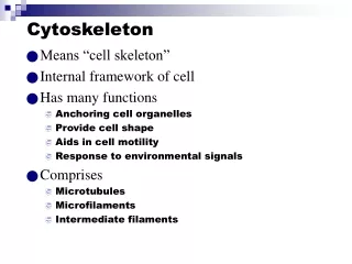
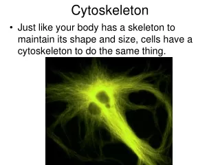
Main functions of cytoskeleton • Determines the shape of the cell • Anchores organelles • Movement of organelles • Tensile strength • Movement of chromosomes • Polarity • Motility
Cytoskeleton • Microfilaments (actin) • Microtubuli (tubulin) • Intermedier filaments • Motor proteins • Actin and microtubule associated proteins
Microfilaments Microtubuli Intermedier filaments
SLIDING Globular proteins Ca2+ ATP Motor proteins Fibrillar proteins
Polymerization of actin + ATP ADP Depolymerization - cytochalasin – inh. phalloidin - stabilizer ATP ADP Pi Polymerization - slow
Actin - still in Prokaryots ! ((Ent et al. Nature 2001,413, 39)
Other actin homologues ((Roeben A et al. J Mol. Biol2006, 358, 145)
Comparison of homologues • Polymerization in both forms • Opposite chirality !!! ((Wickstead and Gull J Cell. Biol2011, 194, 513)
Moving cytoplasm Stationary (cortical) cytoplasm Plasma membrane Actin filaments Cell-wall Chloroplasts Cyclosis • Transitional connections between actin and myosin • Ca2+, temperature- and pH-dependent (Lodish, H. et al. Mol. Cell Biol. 2000, 767)
„Fountain” mechanism Ca2+-dep. requires ATP Mono- Poly- Lobo- podial Filo- Reticulo- Formation of pseudopodium stress-fibrillums integrins
Cross-linking proteins of actin contractile bundle a actinin – in stress fibr. gel-like network filamin - cortex „tight” parallel bundle fimbrin – in filopodium
Migrating keratinocyte 15 mm/sec Formation of lobopodium microtubuli actin-network
- + Regulator proteins of actin polymerisation gCAP39 Severin Gelsolin Villin CapZ Tropomodulin Cofilin Severin Gelsolin
Actin polymerization – acrosomal-reaction (Lodish, H. et al. Mol. Cell Biol. 2000, 767)
local actin polymerization • speed: 10 mm/min • high ability to transmit • in tissues Listeria monocytogenes actin (Fred Soo & Julie TheriotLaboratory
Model of actin nucleation WASP = Wiscott-Aldrich syndr. prot.
Structure of cortical region (Svitkina, TM, Borisy GG J. Cell Biol. 1999, 145, 1009)
Myosin I. Arp2/3 Profilin - G-actin Filamin Integrin Actin – membrane links membrane F-Actin
Profilin-mechanism Tb4 =thymosin b4 Proline-rich protein (Lodish, H. et al. Mol. Cell Biol. 2000, 767)
Filamin – Membrane link filamin actin
Structure of focal contact actin filament a actinin vinculin + paxillin talin integrin fibronectin
Thrombocyte Glycophorin Ankyrin Spectrin tetramer Muscle Epithel A plasma membrane – cortex links ((Lux SE, 1979 Nature 281:426)
E Electromagnetic field induces the transformation of cytoskeleton and formation of pseudopodia Adhesion plaque + + + - -
ATP - ADP Pi Myosin head Ca2+-dependent phosphorylation and its effect on the 3D strcture light chain heavy chain a helix myosin I. 150 kD monomer myosin I I. 260 kD Head: - ATP-ase - motor dimer
Distribution of myosines in the migrating Dyctiosteliumand in dividing cell myosin I. (green) myosin II. (red) (Fukui, Y. Mol. Cell Biol 2000, 785))
+ - Main types of interactions between the globular and fibrillar components of cytoskeleton membrane
MT-blocked F-actin blocked Non-treated
Tubulin – still in Prokaryotes ! FtsZ Tubulin (Margolin Laboratory, University of Texas)
Comparison of homologues • Polymerization in both forms • Monomers build helical structure vs. dimers build tubulus ((Wickstead and Gull J Cell. Biol2011, 194, 513)
Polymerization of tubulin GTP Polymerization - fast GTP GTP GTP Protofilament (strait) GDP GDP GDP GDP Protofilament (curved) Depolymerization
Nucleation Elongation Dynamics of microtubule-assembly - + incorporation balanced release
Role of g-tubulin in nucleation (Wiease et al. Curr.Opin.Struct.Biol. 1999, 9, 250)
Interphase cell centrosome Cilla Basal body Dividing cell spindle Neuron centrosome axon Microtubular systems in the cells -Centrosome - Cilia / flagellum - Mitotic system - Vesicular transport
specific region of the cortex MTOC = Microtubulus organizing center g-tubulin ((Brinkley, B.R. Encyclop. Neurosci. 1987, 665)
24 nm a-bdimer Protofilaments alphatubulin betatubulin Network of microtubuli Fibroblast
Cilia cilia flagellum Paramecium
tubulin (13 ill. 11 protofilaments) A B dynein-arms nexin
The arm moves toward the - pole Composition of dynein-arms ATP-independent binding ATP-dependent hydrolysis
The role of dynein arms in beating of cilia Bending „Telescoping” Proteolysis
Molecules composing the cilia more than 250 types of molecules • 70% a and b tubulin • dynein arms • outer - 9 polypeptides - ATP-ase • inner – composition varies • radial spokes - 17 polypeptides
intermedier filament i.e. vimentin microtubule = rupture actin filament Mechanical characterization of cytoskeleton components deformation force
Role of intermedier filaments Buffer against external mechanical stress Tissue specificity Nucleus – lamines (lamina fibrosa) Epithel – keratin Connective tissue Muscles Neuroglia Neurones - neurofilaments }vimentin
Structure of intermedier filamentums (Lodish, H. et al. Mol. Cell Biol. 2000, 767)