Download
1 / 38
380 likes | 541 Views
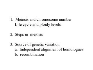
1. Meiosis and chromosome number Life cycle and ploidy levels Steps in meiosis Source of genetic variation Independent alignment of homologues b. recombination. Gametes have a single set of chromosomes. Gametes are haploid, with only one set of chromosomes Somatic cells are diploid.

E N D
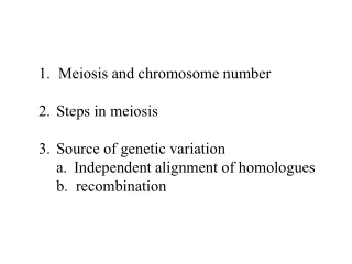
1. Meiosis and chromosome number • Life cycle and ploidy levels • Steps in meiosis • Source of genetic variation • Independent alignment of homologues b. recombination
Gametes have a single set of chromosomes • Gametes are haploid, with only one set of chromosomes • Somatic cells are diploid.
Haploid gametes (n = 23) Egg cell • The human life cycle • Meiosis creates gametes • Mitosis of the zygote produces adult bodies Sperm cell MEIOSIS FERTILIZATION Diploidzygote (2n = 46) Multicellulardiploid adults (2n = 46) Mitosis anddevelopment Figure 8.13
Meiosis reduces the chromosome number from diploid to haploid • Chromosomes are duplicated before meiosis, then the cell divides twice to form four daughter cells.
MEIOSIS I: Homologous chromosomes separate INTERPHASE PROPHASE I METAPHASE I ANAPHASE I Centrosomes(withcentriolepairs) Microtubules attached tokinetochore Metaphaseplate Sister chromatidsremain attached Sites of crossing over Spindle Nuclearenvelope Sisterchromatids Tetrad Centromere(with kinetochore) Homologouschromosomes separate Chromatin Figure 8.14, part 1
While paired, they cross over and exchange genetic information • homologous pairs are then separated, and two daughter cells are produced • In meiosis I, homologous chromosomes are paired
MEIOSIS II: Sister chromatids separate TELOPHASE IAND CYTOKINESIS TELOPHASE IIAND CYTOKINESIS PROPHASE II METAPHASE II ANAPHASE II Cleavagefurrow Sister chromatidsseparate Haploiddaughter cellsforming Figure 8.14, part 2
sister chromatids of each chromosome separate • result is four haploid daughter cells • Meiosis II is essentially the same as mitosis
MITOSIS MEIOSIS Diploid Diploid 1 gamete precursor somatic cell 2n 2n duplication 2 2n 2n 3 2n 2n 4 2n 2n division diploid haploid 5 2n 2n 1n 1n 6 division 7 1n 1n 1n 1n
MITOSIS MEIOSIS PARENT CELL(before chromosome replication) Site ofcrossing over MEIOSIS I PROPHASE I Tetrad formedby synapsis of homologous chromosomes PROPHASE Chromosomereplication Chromosomereplication Duplicatedchromosome(two sister chromatids) 2n = 4 Chromosomes align at the metaphase plate Tetradsalign at themetaphase plate METAPHASE I METAPHASE ANAPHASE I TELOPHASE I ANAPHASETELOPHASE Sister chromatidsseparate duringanaphase Homologouschromosomesseparateduringanaphase I;sisterchromatids remain together Haploidn = 2 Daughtercells of meiosis I 2n 2n No further chromosomal replication; sister chromatids separate during anaphase II MEIOSIS II Daughter cellsof mitosis n n n n Daughter cells of meiosis II Figure 8.15
Genetic variation among offspring is a result of 1) Independent orientation of chromosomes in meiosis 2) random fertilization • Each chromosome of a homologous pair comes from a different parent • Each chromosome thus differs at many points from the other member of the pair
POSSIBILITY 1 POSSIBILITY 2 Two equally probable arrangements of chromosomes at metaphase I Metaphase II Gametes Combination 1 Combination 2 Combination 3 Combination 4 Figure 8.16
Homologous chromosomes carry different versions of genes at corresponding loci
Coat-color genes Eye-color genes C E Brown Black C E C E c e c e c e White Pink Tetrad in parent cell(homologous pair ofduplicated chromosomes) Chromosomes ofthe four gametes Figure 8.17A, B
Crossing over further increases genetic variability • Crossing over is the exchange of corresponding segments between two homologous chromosomes • Genetic recombination results from crossing over during prophase I of meiosis, which increases variation further
Tetrad Chaisma Centromere Figure 8.18A
Coat-colorgenes Eye-colorgenes Tetrad(homologous pair ofchromosomes in synapsis) 1 Breakage of homologous chromatids • How crossing over leads to genetic recombination 2 Joining of homologous chromatids Chiasma Separation of homologouschromosomes at anaphase I 3 Separation of chromatids atanaphase II and completion of meiosis 4 Parental type of chromosome Recombinant chromosome Recombinant chromosome Parental type of chromosome Figure 8.18B Gametes of four genetic types
MEIOSIS I PROPHASE I METAPHASE I ANAPHASE I END OF INTERPHASE
MEIOSIS METAPHASE II TELOPHASE I PROPHASE II ANAPHASE II TELOPHASE II
INDEPENDENT ASSORTMENT TELOPHASE II METAPHASE II METAPHASE I METAPHASE I
SPERMATOGENESIS b OOGENESIS a spermatogonium oogonium primary spermatocyte primary oocyte meiosis l secondary spermatocyte secondary oocyte polar body meiosis ll spermatids polar bodies (will be degraded) egg
8.21 Accidents during meiosis can alter chromosome number Nondisjunctionin meiosis I • Abnormal chromosome count is a result of nondisjunction • Either homologous pairs fail to separate during meiosis I Normalmeiosis II Gametes n + 1 n + 1 n – 1 n – 1 Number of chromosomes Figure 8.21A
Or sister chromatids fail to separate during meiosis II Normalmeiosis I Nondisjunctionin meiosis II Gametes n + 1 n – 1 n n Number of chromosomes Figure 8.21B
Fertilization after nondisjunction in the mother results in a zygote with an extra chromosome Eggcell n + 1 Zygote2n + 1 Spermcell n (normal) Figure 8.21C
8.20 Connection: An extra copy of chromosome 21 causes Down syndrome • This karyotype shows three number 21 chromosomes • An extra copy of chromosome 21 causes Down syndrome Figure 8.20A, B
The chance of having a Down syndrome child goes up with maternal age Figure 8.20C
8.22 Connection: Abnormal numbers of sex chromosomes do not usually affect survival • Nondisjunction can also produce gametes with extra or missing sex chromosomes • Unusual numbers of sex chromosomes upset the genetic balance less than an unusual number of autosomes
8.23 Connection: Alterations of chromosome structure can cause birth defects and cancer • Chromosome breakage can lead to rearrangements that can produce genetic disorders or cancer • Four types of rearrangement are deletion, duplication, inversion, and translocation
Deletion Duplication Homologouschromosomes Inversion Reciprocaltranslocation Nonhomologouschromosomes Figure 8.23A, B
Translocation Figure 8.23Bx
A chromosomal translocation in the bone marrow is associated with chronic myelogenous leukemia • Chromosomal changes in a somatic cell can cause cancer Chromosome 9 Reciprocaltranslocation Chromosome 22 “Philadelphia chromosome” Figure 8.23C Activated cancer-causing gene