Download
1 / 4
80 likes | 627 Views
Fig 39. Tetralogy of Fallot. Fig 40. Tetralogy of Fallot. RVOT. RV. VSD. Ao. LV. Ao. Parasternal long axis view showing aortic overriding and large sub-aortic VSD (arrow) in a patient with Tetrology of Fallot. . Parasternal short axis view demonstrating

E N D
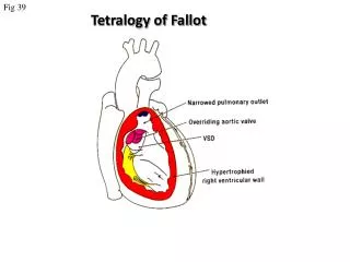
Fig 39 Tetralogy of Fallot
Fig 40 Tetralogy of Fallot RVOT RV VSD Ao LV Ao Parasternal long axis view showing aortic overriding and large sub-aortic VSD (arrow) in a patient with Tetrology of Fallot. Parasternal short axis view demonstrating The right ventricular outflow tract is narrowed by the outlet septum.
Fig 41 Tetralogy of Fallot RVOT Ao Continuous wave Doppler trace taken from the RVOT showing the peak velocity of more than 4 m/s. Colour Doppler showing a narrowed right ventricular outflow tract (RVOT) with turbulent flow.
Fig 42 Repaired Fallot LV RV RVOT LA Ao RA RA Parasternal short axis view showing a residual right ventricular outflow tract obstruction in an adult patient with repaired Tetralogy of Fallot Apical four chamber view demonstrating a severely dilated RA from long standing high right ventricular pressure and tricuspid regurgitation.