Effects of Exercise on Neural Activity in Antisaccade Tasks Among Overweight Children: An fMRI Study
This study investigates how exercise influences neural substrates involved in antisaccade performance in overweight children. We analyzed brain activity patterns through fMRI while engaging children in antisaccade tasks, revealing differences between those who participated in an eight-week exercise program and those who did not. Findings indicate that the exercise group showed increased activity in the prefrontal cortex and decreased activity in regions associated with saccadic movements. This suggests enhanced cognitive efficiency and automaticity in task performance due to exercise.

Effects of Exercise on Neural Activity in Antisaccade Tasks Among Overweight Children: An fMRI Study
E N D
Presentation Transcript
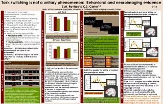
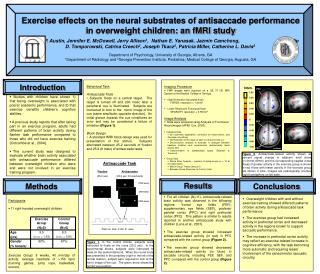
Exercise effects on the neural substrates of antisaccade performance in overweight children: an fMRI study Benjamin P. Austin, Jennifer E. McDowell, Jerry Allison1, Nathan E. Yanasak, Jazmin Camchong, Phillip D. Tomporowski, Catrina Creech2, Joseph Tkacz2, Patricia Miller, Catherine L. Davis2 Department of Psychology, University of Georgia, Athens, GA 1Department of Radiology and 2Georgia Prevention Institute, Pediatrics; Medical College of Georgia, Augusta, GA • Introduction • Studies with children have shown 1) that being overweight is associated with poorer academic performance, and 2) that exercise benefits children’s cognitive abilities. • A previous study reports that after taking part in an exercise program, adults had different patterns of brain activity during flanker task performance compared to those who did not have exercise training (Colcombe et al., 2004). • The current study was designed to evaluate whether brain activity associated with antisaccade performance differed between overweight children who were and were not involved in an exercise training program. Behavioral Task Antisaccade Trials • Subjects fixate on a central target. The target is turned off and 200 msec later a peripheral cue is illuminated. Subjects are instructed to look to the mirror image of the cue (same amplitude, opposite direction). An initial glance towards the cue constitutes an error and may be considered a failure of inhibition (Figure 1). • Block Design • A standard fMRI block design was used for presentation of the stimuli. Subjects alternated between 25.2 seconds of fixation and 25.2 (8 trials) of antisaccade tasks. Imaging Procedure • FMR images were acquired on a GE 3T HD MRI System at the Medical College of Georgia. • High Resolution Structural Scan FSPGR; resolution = 1.3mm3 • Lower Resolution Functional Scan SPGR-EPI; resolution = 3.75mm3 • Image Analysis • Data were analyzed using Analysis of Functional NeuroImages (AFNI; Cox, 2003). • Individual Data • 3D volumetric registration, correction for head motion, and Gaussian smoothing • Normalization to the mean of each voxel across the run • Deconvolution analysis to evaluate % changes between baseline (fixation) and experimental (antisaccade) tasks across time • Transformation to standardized space (Talaraich & Tournoux) • Group Data • Whole Brain Analysis – clusters of active voxels (n > 12 at 0.5 alpha level) • Within Group T-tests (versus 0) • Between Group (Exercise vs Control) t-test Inten. PFC 1.00 0.80 0.60 0.40 PPC 0.20 z=41mm 0 SEF -0.20 FEF -0.40 -0.60 -0.80 -1.00 PPC z=45mm Figure 2. Antisaccade-related activity shown as percent signal change in adjacent axial slices (z=41mm,45mm) and the corresponding sagittal views. Areas of greater activity in the exercise group is shown in red, those with lesser activity in the exercise group are shown in blue. Images are radiologically oriented (right hemisphere on left side). Antisaccade Task Fixation (25.2 sec) Antisaccades (25.2 sec; 8 trials/block) Results Methods Participants • 11 right-handed overweight children Exercise Group: 8 weeks, 40 min/day of activity, average heartrate of >150 bpm (running games, jump rope, basketball, soccer). Conclusions (1700 msec) (200 msec) • For all children (N=11), antisaccade-related brain activity was observed in the following regions: frontal eye fields (FEF), supplementary eye fields (SEF), posterior parietal cortex (PPC) and right prefrontal cortex (PFC). This pattern is similar to results reported in another antisaccade study with children (Luna et al., 2001). • The exercise group showed increased antisaccade-related activity (in red) in PFC compared with the control group (Figure 2). • The exercise group showed decreased antisaccade-related activity (in blue) in saccade circuitry, including FEF, SEF, and PPC compared with the control group (Figure 2). • Overweight children with and without exercise training showed different patterns of brain activity during antisaccade task performance. • The exercise group had increased activity in prefrontal cortex and decreased activity in the regions known to support saccadic performance. • The increase in prefrontal cortex activity may reflect an exercise-related increase in cognitive efficiency, with the task becoming more automatic and requiring lesser involvement of the sensorimotor saccadic circuitry. (1250 msec) Total run time: 5 min 31 secs Figure 1. In the control blocks, subjects were instructed to fixate on the cross (25.2 sec). In the experimental blocks, subjects were instructed to fixate on the central diamond. When the round circle was presented in the periphery (right or the left of the central fixation), subject were required to look at the mirror image of the cue. The green arrow shows the correct eye position.