Download
1 / 28
280 likes | 583 Views
The Chemical Senses: Gustatory, Visceral Afferents and Olfactory Systems. I. Gustatory and Visceral Afferent Systems. Overview of the systems. Cranial nerve branches and sensory organs for taste and visceral afferent system.

E N D
The Chemical Senses: Gustatory, Visceral Afferents and Olfactory Systems
I. Gustatory and Visceral Afferent Systems • Overview of the systems. • Cranial nerve branches and sensory organs for taste and visceral afferent system. • Solitary nuclear complex and its projections for gustatory system and visceral afferents. • Thalamic projections for gustatory system: parvocellular portion of the VPM. • 1° cortical gustatory areas: Frontal operculum and the anterior insular cortex.
II. Olfactory System • Overview and special features of this system – direct projection with thalamic relay; - 1° cortex = allocortex. • 1° olfactory neurons in nasal mucosa. • Olfactory bulb. • 1° olfactory cortex constitutes 5 different brain areas: • Anterior olfactory nucleus. • Amygdala. • Olfactory tubercle. • Piriform/periamygdaloid cortices. • Entorhinal cortex.
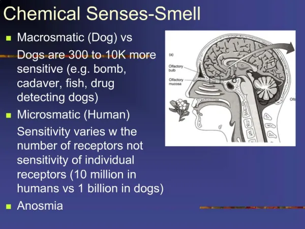
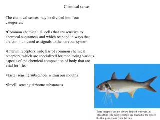
Specialized neurons present in the olfactory epithelium in the nose. They project cilia into a mucus layer. The cilia are able to bind to odorant molecules the binding triggers an AP which is transmitted to the olfactory area of the olfactory bulb olfactory cortex (lower frontal area and limbic system of the brain Each olfactory receptor is specialized for 1 odorant molecule Olfaction
I. Gustatory and Visceral Afferent Systems • Overview of the systems. Taste is mediated by 3 different cranial nerves: facial (VII), glossapharyngeal (IX), and vagus (X). Visceral afferent system uses largely IX and X.
In the gustatory system, receptor cells (“taste buds”) are separate from the 1° afferent fibres. This is an e.g. where the job of transducing the info and transmitting it are done by separate cells. 1° afferent fibres from both systems collect in the solitary tract in the rostral medulla, and then terminate in the solitary nucleus. The gustatory portion of this nucleus is more rostral than the cardiorespiratory portion.
Receptors for taste are modified epithelial cell present in taste buds located on the tongue, roof of the mouth and pharynx Taste
Ascending Gustatory Pathway • Gustatory fibres ascend • ipsilaterally in the central • tegmental tract Medial portion of VPN 1° cortex (frontal operculum and anterior insular cortex) – Mediates the discriminative aspects of taste.
Four primary types of taste receptors : sour, salt, sweet and bitter (and a new one: umami) • The binding of the receptor to a taste molecule triggers the entry of calcium in the cell release of neurotransmitter in a synapse with a neuron
Neural pathway • Taste impulses travel through nerves VII, IX and X to a gustatory nucleus in the medulla oblongata (cross over) thalamus gustatory cortex located in the parietal lobe in the mouth area.
Ascending Visceral Afferent System Ascending projections synapse in the parabrachial nucleus of the pons VPM Amygdala and hypothalamus – For regulating visceral and food intake.
I. Gustatory and Visceral Afferent Systems B. Cranial nerve branches and sensory organs. • Taste (gustatory system) Taste buds contain the receptor cells which transduce soluble chemical stimuli into a neural signal. These have synaptic contact with 1° afferent fibres. On tongue: clustered in papillae (Fig. 9-2B): anterior 2/3 branch of VII (facial) posterior 1/3 IX (glossopharyngeal) Also palate VII epiglottis, larynx X pharynx IX
I. Gustatory and Visceral Afferent Systems B. Cranial nerve branches and sensory organs. 2. Visceral afferents IX, X also have branches which serve arterial bp receptors in carotid sinus, aortic arch. X respiratory structures, GIT (rostral to splenic flexture) – pseudounipolar neurons, much like 1° afferents for somatic sensory system. Both systems come together in the ganglion located just outside the brainstem (Fig. 9-3).
Fig. 9-3: VII geniculate ganglion; IX, X inferior ganglion (or petrosal, nodose) rostral medulla
C. Solitary Nuclear Complex • Gustatory separation within • bs solitary tract • surrounding solitary nucleus • (anterior portion) ipsilateral • central tegmented tract • thalamus. • 2. Cardiorespiratory nucleus • (caudal portion of solitary complex. • Receives a variety of inputs from • CV, respiratory, and GIT systems • “internal state” descending (controls HR, bp, gut motility, secretions, etc.) Ascending parabrachial n. within pons amy, hypo {integrates visceral info with autonomic functions}
E. Primary Gustatory Cortex (Fig. 9-6) Frontal operculum and anterior insular cortex: Modalities of touch and taste from the tongue are represented separately
II. Olfactory System • Overview + special features of nerve = cranial nerve I: • Info goes to 1° cortex with thalamic relay. • 1° olfactory cortex = phylogenetically older allocortex. • Projections to 5 separate cortical regions, rather than to just 1 (all allocortex) (Fig. 9-7, below): - less developed in humans, compared to some other species. - less precise topographic organization than other systems we have reviewed.
II. Olfactory System B. 1° olfactory neurons in nasal mucosa (olfactory mucosa) in superior nasal concha contains bipolar neurons which are chemosensitive small fasciculus cribiform plate of ethmoid bone olfactory nerve) -Note risk of anosmia with head trauma (shearing of these fibres. Receptor cells have olfactory cilia (like many other sensory systems), containing the machinery for receiving chemical stimuli (multiple types of transmembrane receptors recognize molecular characteristics of oderants).
II. Olfactory System C. Olfactory bulb – 1st CNS relay for olfactory input: - on ventral surface of brain - neurons organized into discrete layers Fig. 9-7
Fig. 9-8. Input to neurons with discrete morphological units, called Glomeruli (surrounded by glial sheath) – limits the spread of neurotransmitters, neurons, mitral and tufted cells (olfactory tract) Other neurons receiving inputs: periglomular cells (input directly from olfactory nerve) and granule cells (receives excitatory input from mitral cells). Both of these cells are inhibitory interneurons, providing feedback.
II. Olfactory System Projections (Fig. 9-9): Olfactory tract bifurcates into lateral and medial olfactory striae. Axons from other brain regions projecting to olfactory bulb synapse with the medial. Axons from the olfactory bulb itself lateral. One 1° projection (cortical region) is seen directly caudal to these on basal forebrain: olfactory tubercle (location in a region called the anterior perforated substance)
D. Primary olfactory cortical areas(Fig. 9-7) Most areas are alocortex (more specifically, 3-layered paleocortex – archicortex associated with hippocampus)
D. Primary olfactory cortical areas • Anterior olfactory nucleus – modulates info processing in the olfactory bulb. • Amygdala – in ant temporal lobe for self-regulation of various behaviors (e.g., feeding (endocrine), reproductive behaviors), via projection to hypothalamus. • Olfactory tubercle – part of basal forebrain projections to and from olfactory bulb – play a role in regulating emotion. • Piriform + periamygdaloid cortices – ant temporal lobe (shaped like a pear): olfactory perception as in: - input in internal processing of odors. - projects to frontal neocortical areas for olfactory discrimination (in part, through thalamus). 5. Rostral entorhinal cortex – on parahippocampal gyrus. - projects to hippocampus - modulates the association of odors with long-term memories.