Download
1 / 85
850 likes | 888 Views
Learn to recognize, label, and identify structures in functional and immature testes on slides of mature dog testis. Explore spermatogenesis stages and Leydig cell function. Educational resource for students.

E N D
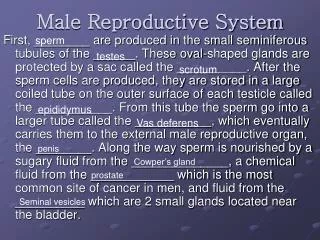
MALE REPRODUCTIVE This resource is licensed under the Creative Commons Attribution Non-Commercial & No Derivative Works License
Objectives Students should be able to: Recognise, draw and label the main structures of a section of functional testis and immature testis. Identify the levels of the epididymis according to their cellular structural variations. Label the regions of the spermatozoon.
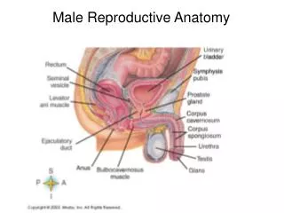
DEMO SLIDE Mature testis 5.0 mm Section of whole testis : Identify the main features of the section.
DEMO SLIDE Mature testis E : epididymis R : rete testis mediastinum testis ductus deferens E E 5.0 mm R Section of whole testis : Identify the main features of the section. tunica albuginea E connective tissue septa dividing the testis into lobules seminiferous tubules
SLIDE 162 Mature testis (dog) At low magnification identify : tunica albuginea lobules connective tissue septa seminiferous tubules 1.0 mm
SLIDE 162 Mature testis (dog) connective tissue septa lobule At low magnification identify : tunica albuginea lobules connective tissue septa seminiferous tubules Septa extend from the tunica albuginea across the testis dividing it into lobules. tunica albuginia seminiferous tubules 1.0 mm
SLIDE 162 Mature testis (dog) The lobules are composed of convoluted seminiferous tubules. Lying between these tubules in the interstitial tissue are found Leydig cells (endocrine cells). 250 µm
SLIDE 162 Mature testis (dog) ST : seminiferous tubules L : area of Leydig cells ST The lobules are composed of convoluted seminiferous tubules. Lying between these tubules in the interstitial tissue are found Leydig cells (endocrine cells). connective tissue septum L ST 250 µm
SLIDE 162 Mature testis (dog) What magnification is best for identifying this section? Give reasons. 100 µm
What magnification is best for identifying this section? Give reasons. Higher magnifications are best. It is then possible to distinguish the different cell types present. At low magnification the tissue can be confused with epididymis. SLIDE 162 Mature testis (dog) L ST ST : seminiferous tubules L : Leydig cells What magnification is best for identifying this section? Give reasons. L ST ST L 100 µm
SLIDE 162 Mature testis (dog) What is secreted by the Leydig cells? 50 µm
What is secreted by the Leydig cells? Leydig cells synthesize and secrete testosterone. These cells have a ‘foamy’ appearance due to the presence of lipid droplets and granules. SLIDE 162 Mature testis (dog) C : capillary Leydig cells C What is secreted by the Leydig cells? 50 µm
SLIDE 162 Mature testis (dog) Can you explain the appearance of tubules and cells from your knowledge of the architecture of the organ? 50 µm
Can you explain the appearance of tubules and cells from your knowledge of the architecture of the organ? Seminiferous tubules are surrounded by lymph spaces in the interstitium and Leydig cells. These tubules are lined by Sertoli cells. Germ cells differentiate and mature in compartments between the Sertoli cells. SLIDE 162 Mature testis (dog) L L : Leydig cells Can you explain the appearance of tubules and cells from your knowledge of the architecture of the organ? L blood capillary L seminiferous tubule 50 µm
SLIDE 162 Mature testis (dog) In sections; adjacent seminiferous tubules show different stages of spermatogenesis. Tight junctions between Sertoli cells divide the basal and luminal compartments. As germ cells progress from the basal to the luminal compartment they undergo division and differentiation : 50 µm
SLIDE 162 Mature testis (dog) SPERMATOGONIA ↓ PRIMARY SPERMATOCYTES ↓ SECONDARY SPERMATOCYTES ↓ SPERMATIDS ↓ SPERMATOZOA In sections; adjacent seminiferous tubules show different stages of spermatogenesis. Tight junctions between Sertoli cells divide the basal and luminal compartments. As germ cells progress from the basal to the luminal compartment they undergo division and differentiation : 50 µm
SLIDE 162 Mature testis (dog) Examine the tubules at higher power (X40). Are they structurally identical? 25 µm
Examine the tubules at higher power (X40). Are they structurally identical? No, adjacent tubules are at different stages of spermatogenesis. Tubules also vary in the angle at which they have been sectioned (not all true TS). SLIDE 162 Mature testis (dog) P : primary spermatocytes ES : elongated spermatids Examine the tubules at higher power (X40). Are they structurally identical? spherical spermatids ES spermatogonia P nucleus of Sertoli cell P P 25 µm
SLIDE 162 Mature testis (dog) Describe what you see. 25 µm
Describe what you see. In this tubule many primary spermatocytes are visible. Many spherical spermatids are seen in the mid regions and elongated spermatids with tails protruding into the lumen towards the apical margins of the Sertoli cells. A prominent myoid or myoepithelial cell can be seen outside the tubule. SLIDE 162 Mature testis (dog) M : myoid (myoepithelial) cell Describe what you see. elongated spermatids spherical spermatids primary spermatocytes 25 µm M
SLIDE 162 Mature testis (dog) Describe what you see. 25 µm
Describe what you see. Prominent in this tubule are spherical spermatids many of which are in the cap phase. Note also sperm tails in the lumen of the tubule. SLIDE 162 Mature testis (dog) S : spermatogonia P : primary spermatocytes S Describe what you see. spherical spermatids with acrosomal caps P P S 25 µm
SLIDE 162 Mature testis (dog) Describe what you see. 25 µm
Describe what you see. This section of tubule has both prominent spherical spermatids in the golgi phase and cap spermatids. The elongated spermatids are still enveloped by the apical portions of the Sertoli cells. SLIDE 162 Mature testis (dog) S : spermatogonia P : primary spermatocytes Si : spherical spermatids Sii : cap spermatids L : Leydig cells P L Si S Describe what you see. Sii sperm tails Si P Sii S 25 µm
SLIDE 162 Mature testis (dog) Describe what you see. 25 µm
SLIDE 162 Mature testis (dog) Describe what you see. Again most of the spherical spermatids are in the acrosomal cap phase. The darker strands of tissue are the bodies of the Sertoli cells. SLIDE 162 Mature testis (dog) Sertoli cells acrosomal cap spermatids 25 µm
TRANSMISSION ELECTRON MICROGRAPH OF DOG SPERMATOZOA head containing nucleus covered by acrosome head neck mitochondria mid-piece axial filaments 9+2 tail 2.5 µm
SCANNING ELECTRON MICROGRAPH OF DOG SPERMATOZOA A group of spermatozoa on the surface of the uterine horn. Note the surface processes of the uterine epithelial cells. 10 µm
SLIDE 163 Immature testis (ferret) Low magnification showing main body surrounded by tunica albuginea. At the top is the epididymis. 200 µm
SLIDE 163 Immature testis (ferret) epididymis Low magnification showing main body surrounded by tunica albuginea. At the top is the epididymis. tunica albuginea developing tubules 200 µm
SLIDE 163 Immature testis (ferret) Compare with previous slide 162. 250 µm
SLIDE 163 Immature testis (ferret) tubules blood vessels Compare with previous slide 162. tunica albuginea 250 µm
SLIDE 163 Immature testis (ferret) List the similarities and differences. 100 µm
List the similarities and differences. Similarities. Both have tubules. Leydig cells present, but function at low level. SLIDE 163 Immature testis (ferret) 100 µm
List the similarities and differences. Similarities. Differences. Both have tubules. Closely packed tubules, little or no lumen. Leydig cells present, but function at low level. No spermatogenesis. Immature Sertoli cells. Giant gonocytes present. SLIDE 163 Immature testis (ferret) 100 µm
SLIDE 163 Immature testis (ferret) What cell types indicated below are evident in the tubular sections? 50 µm
What cell types indicated below are evident in the tubular sections? Gonocytes (pre-spermatogonia). Sertoli cells. SLIDE 163 Immature testis (ferret) G : gonocytes What cell types indicated below are evident in the tubular sections? G G Sertoli cells G G 50 µm
SLIDE 163 Immature testis (ferret) Higher magnification of developing germ cells, Gonocytes within the tubules. These will become spermatogonia. Leydig cells develop in the interstitial areas. 25 μm
SLIDE 163 Immature testis (ferret) I : interstitial tissue site of Leydig cells G : gonocytes within tubules G capillaries Higher magnification of developing germ cells, Gonocytes within the tubules. These will become spermatogonia. Leydig cells develop in the interstitial areas. I Sertoli cells forming the epithelial lining of tubules I G 25 μm
DEMO SLIDE Mature testis Section of whole testis : Identify the regions of the epididymis. 5.0 mm
DEMO SLIDE Mature testis A : head B : body C : tail B B Section of whole testis : Identify the regions of the epididymis. Efferent ductules lead from the rete testis and join to form the head of the epididymis. C A 5.0 mm
DEMO SLIDE Mature testis At low magnification identify the main features of the section. 2.5 mm
DEMO SLIDE Mature testis body of epididymis (corpus) tail of epididymis (cauda) At low magnification identify the main features of the section. tunica albuginea tubules of testis 2.5 mm
SLIDE 166 Epididymis What might this organ be confused with? 1.0 mm
What might this organ be confused with? Testis. Differential vesicular glands, bulbourethral gland, prostate gland. At higher magnification with kidney tubules. SLIDE 166 Epididymis head (caput) What might this organ be confused with? body (corpus) tail (cauda) 1.0 mm
SLIDE 166 Epididymis What distinguishing features can you identify? 100 µm
What distinguishing features can you identify? Pseudostratified epithelium with stereocilia. No interstitial cells. Spermatozoa in lumen of duct. SLIDE 166 Epididymis spermatozoa in lumen What distinguishing features can you identify? pseudostratified columnar epithelium (principal cells) and smaller basal cells forming wall of tubules 100 µm
SLIDE 166 Epididymis What is the function of the cellular extensions projecting into the lumen? 25 µm
What is the function of the cellular extensions projecting into the lumen? These are stereocilia (long branching microvilli). They are often clumped together and indicate a resorptive function of these cells. SLIDE 166 Epididymis spermatozoa What is the function of the cellular extensions projecting into the lumen? stereocilia (clumped) pseudostratified epithelium columnar cells basal cells 25 µm