Download
1 / 58
600 likes | 636 Views
This case study presents an 80-year-old male with myelodysplastic syndromes, prostate cancer, gout, OSA, and CAD. Laboratory data, clinical presentation, physical exam findings, and bone marrow biopsy results provide insight into his complex medical condition. The text explores the diagnosis, treatment options, and challenges associated with managing this patient's overlapping health issues.

E N D
80 yo male: OSA, gout, CAD, and prostate cancer who was referred for progressive anemia. • Past few months: noticed his health started to decline. Localized prostate cancer: end of 2005, tx external beam radiation. • Prior to any radiation: wbc 4.1 Hg 9.5 Plt 193 and MCV of 102. • One month after radiation: Hg 8.6 and plt 138. Progressive weakness and DOE. No chest pain. No GIB. He received 2 units of prbcs while waiting for results of work-up.
A week later: shortness of breath, worsening DOE, and generalized weakness. • His hemoglobin dropped down to 8.2 from 10. • Admitted to the hospital: found to have a non-ST elevation myocardial infarction. • Underwent a cardiac catherization which showed a 3-vessel disease. • CABG was recommended. Path of the bone marrow biopsy showed MDS. • Question: Should this patient undergo a CABG?
PMH: OSA on CPAP; Gout • CAD: diagnosed 30 years ago; told to have blockage • Prostate cancer: diagnosed Dec 2005 undergoing radiation • Social: widow, 60 pack year, quit 30 years ago; occasional etoh; former lawyer • Meds: Lortab, Vytorin, Toprol XL, Digitek
Pex: appears tired, DOE from door to room • No LAD, no oral lesions • Chest: bilateral rales about ¼ up • CV: regular RR with 3/6 murmur • Abd: unremarkable • Ext: 1+ edema of LLE to knee • Neuro: non-focal, alert, oriented
Laboratory data: • Wbc 4.1 hg 8.9 hct 26.7 plt 158 mcv 103 ANC 3321 • Creatinine 1.2 Folate >24 Ferritin 222 B12 352 • Reticulocyte count 3.4 with an absolute of 84K • Serum erythropoeitin level 498
Bone marrow biopsy • Cellularity: 70-75%, increased for patient’s age • Myeloid: adequate, immature cells/blasts not increased • Megakaryocytes: many atypical megakaryocytes noted; mononuclear forms and micromegakaryocytes • Erythroid precursors: dyspoietic changes are present in up to 10% of erythroid series • Flow cytometry: normal, blasts not increased • Cytogenetics: normal
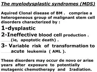
Introduction • MDS: disorders of hematopoietic stem cells • Characterized by dysplastic and ineffective blood cell production • Peripheral blood cytopenias • Variable risk of transformation to acute leukemia • Arise de novo or years after exposure to mutagenic therapy (radiation or chemotherapy)
Clinical Presentation • Signs and symptoms are non-specific • Many asymptomatic • Routine laboratory screening • Others present with sxs from anemia • Fatigue, weakness, angina, dizziness • Infection, easy bruising, bleeding: less common • Fever and wt loss: uncommon
Clinical presentation • Infection: while neutropenia is largely responsible for the high incidence of infection, granulocyte dysfunction may contribute • Bacterial infections predominate • Skin is most common • Infection: principal cause of death in pts with MDS • Fungal, viral, and mycobacterial infx can occur, rare without concurrent immunosuppressive agents
Physical Exam • Hepatomegaly, splenomegaly, LAD: uncommon • Except CMML • Cutaneous manifestations: uncommon • Sweet’s syndrome( neutrophilic dermatosis): transformation to acute leukemia ( IL-6) • Granulocytic sarcoma (chloroma): herald disease transformation into acute leukemia
Laboratory findings • Quantitative changes of one or more blood and bone marrow elements are the hallmark of the disease • Red cell series • Granulocytic series • Platelets • Bone marrow findings
Red cell series • Anemia is almost uniformly present • Inappropriately low reticulocyte response • Anemia may be the only cytopenia or be accompanied by thrombocytopenia or neutropenia • Less than 5% present with an isolated neutropenia or thrombocytopenia in the absence of anemia
Red cell series • Erythrocyte morphology: normocytic or macrocytic • Peripheral smear: basophilic stippling, Howell-Jolly bodies, and nucleated red cells • Ringed sideroblasts: iron granules occupy more than 1/3 of the nuclear rim
Granulocytic series • Leukopenia: found in approximately 50% at time of diagnosis • Granulocytes: display reduced segmentation, aka pseudo-Pelger-Huet • Often with reduced or absent granulation
Platelets • Thrombocytopenia: varying degrees are present in ~25% of pts with MDS • Rarely represent an isolated early manifestation of the disease • Giant plts or circulating megakaryocyte fragments may be present • May have increased bleeding tendency despite adequate plt number
Bone marrow alterations • BM: usually hypercellular • Accompanied by single or multilineage dysplasia • Hypercellular BM/pancytopenia: reflects premature cell loss via intramedullary cell death • Hypocellular: rare • See in therapy-related MDS • Megakaryocytes are normal or increased in number
Auer rods • Within leukemic blasts • FAB: belongs to category of RAEB-T • WHO: its presence should lead to the suspicion that patient has already transformed into AML
Diagnosis • Considered in any patient with unexplained cytopenia(s) • Nuclear hyposegmentation of granulocytes (Huet anomaly) • Mononuclear megakaryocytes, micromegakaryocytes, or megakaryocytes with dysplastic nuclei • Hypogranular neutrophils • Macrocytic or acanthocytic red blood cells • Ring sideroblasts in developing red cell precursors
Differential diagnosis • Above findings not unique to MDS • Exclude other contributing conditions • Alcohol • Megaloblastic anemia • HIV • Medications • Heavy metals
Differential diagnosis • Other causes of macrocytic anemia • B12 and folate deficiency • Megaloblastic anemia have high MCV, reduced retic count, and pancytopenia • Reduced neutrophil lobulation is characteristic of MDS • Combination of increased neutrophil lobulation and macrocytosis is pathognomonic of megaloblastic anemia
Differential diagnosis • HIV: dysplastic hematopoiesis and variable degrees of cytopenia are common findings in HIV • Myelodysplasia was found up to 69% in BM of pts with HIV • Dysplasia can be reversible depending on the cause (meds vs infection) • Medications: associated with acquired dysplastic changes • Valproic acid, mycophenolate, gancyclovir, alemtuzumab • Reversible on reduction or discontinuation of medications
Diagnosis • Need bone marrow biopsy, flow cytometry, immunochemical studies, and cytogenetics • Clonal chromosomal abnormality of 5q- and monosomy 7 confirms the dx of MDS • Deletion 5q
Treatment • Control of symptoms due to cytopenias • Improving quality of life, minimizing toxicity of therapy • Improving overall survival • Decreasing progression to AML
National Comprehensive Cancer Network • Age, performance status, IPSS-defined risk category • Low intensity • Outpatient tx, hematopoietic factors, differentiation-inducing agents, low intensity chemotherapy • High intensity • Intensive combination chemotherapy and hematopoietic cell transplantation • May reduce risk of death from disease
Hematopoietic cell transplantation • Very limited • donor availability, advanced age • Upper age limit for allogeneic HCT ~60 • 75% of pts with MDS >60 • Considered in pts <60, HLA-matched sibling donor, risk of disease progression, excellent PS • Transplant-related mortality and relapse rate at 5 years as high as 40% • Timing of transplantation: development of new cytogenetic abnormality, worsening cytopenia, progression to a higher IPSS group
Chemotherapy • High dose • young • MDS with >10% blasts in BM
Intermediate dose chemotherapy • Azacitidine: pyrimidine nucleoside analog of cytidine • Causes hypomethylation of DNA and direct cytotoxicity on abnormal BM hematopoietic cells • Median time to leukemia was 21 mos vs 13mos • Quality of life was significantly improved • No difference in overall survival • FDA: RA, RARS, RAEB and CMML
Low dose chemotherapy • Immunomodulatory agents • Thalidomide • Lenalidomide • Thalidomide • Low response rate • Adverse side effects: fatigue, constipation, peripheral neuropathy, and drowsiness • Lenalidomide (Revlimid) • Thalidomide derivative without the neurologic toxicity • FDA approved 12/27/05: transfusion-dependent anemia due to low or intermediate-1 risk MDS associated with deletion 5q.
5q- syndrome • Karyotypic and clinical distinctiveness sets it apart from other subtypes • Female predominance (7:3) median age 68 at dx • Transfusion-dependent anemia 80% • Low incidence of neutropenia, thrombocytopenia, infection and bleeding • Low incidence of transformation into acute leukemia (16%) • May respond dramatically to lenalidomide