5.1 Mitochondrial structure and function
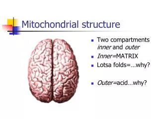
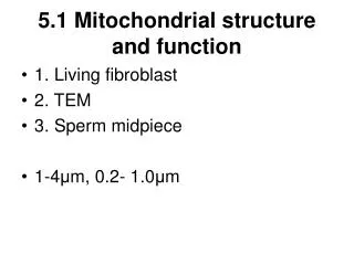
5.1 Mitochondrial structure and function. 1. Living fibroblast 2. TEM 3. Sperm midpiece 1-4 μ m, 0.2- 1.0 μ m. 1. Mitochondrial membranes. The outer membrane is thought to be homologous to an outer membrane present as part of the cell wall of certain bacterial cells.

5.1 Mitochondrial structure and function
E N D
Presentation Transcript
5.1 Mitochondrial structure and function • 1. Living fibroblast • 2. TEM • 3. Sperm midpiece • 1-4μm, 0.2- 1.0μm
1. Mitochondrial membranes • The outer membrane is thought to be homologous to an outer membrane present as part of the cell wall of certain bacterial cells. • The inner membrane is highly impermeable; all molecules and ions require special membrane transporters to gain entrance to the matrix.
2. The mitochondrial matrix • Possess ribosomes, circular DNA to manufacture their own RNAs and proteins
1. The Tricarboxylic Acid (TCA) cycle • Acetyl CoA + 2 H2O + FAD + 3 NAD+ + GDP + Pi→ 2 CO2 + FADH2 + 3 NADH + 3H+ + GTP +HS-CoA
The glycerol phosphate shuttle • Electrons are transferred from NADH to dihydroxyacetone phosphate (DHAP) to form glycerol 3-phosphate, which shuttles them into the mitochondrion. These electrons then reduce FAD at the inner membrane, forming FADH2 which can transfer the electrons to a carrier of the electron-transport chain.
2. The importance of reduced coenzymes in the formation of ATP (Chemiosmosis) • 1. High-energy electrons are passed from FADH2 or NADH to the first of a series of electron carriers in the electron transport chain. • 2. The controlled movement of protons back across the membrane through an ATP-synthesizing enzyme provides the energy required to form ATP from ADP.
5.3 The role of mitochondria in the formation of ATP • 1. Oxidation-reduction potentials • 2. Electron transport • 3. Types of electron carriers
the greater the affinity, the stronger the oxidizing agent. Oxidation –reduction potential Oxidizing agents can be ranked in a series according to their affinity for electrons:
Reducing agents can also be ranked according to their affinity for electrons: • The lower the affinity, the stronger the reducing agent • Reducing agents are ranked according to electron-transfer potential, such as NADH is strong reducing agent, whereas those with low electron-transfer potential such as H2O, are weak reducing agents.
Oxidizing and reducing agents occur as couples such as NAD+ and NADH which differ in their electrons.Strong reducing agents are coupled to weak oxidizing agents and vice versa. For example, in NAD+ - NADH, NAD + is a weak oxidizing agent, in O2– H2O, O2 is a strong oxidizing agent
The affinity of substances for electrons can be measured by instruments that detect voltage—oxidation-reduction (redox) potential.
2. Electron transport • 1. Five of the nine reactions in Fig. 5.7 are catalyzed by dehydrogenases that transfer pairs of electrons fron substrates to coenzymes, NADH and FADH2→ electron-transport chain • 2. NADH and FADH2 dehydrogenase are located in theinner membrane of mitochondria.
3. Types of electron carriers • Flavoproteins (FMN of NADH dehydrogenase) • Cytochromes (heme group) • Three copper atoms • Ubiquinone (Coenzyme Q) • Iron-sulfur proteins • With the exception of ubiquinone, all of the redox centers within the respiratory chain that accept and donate electrons are prosthetic groups (non-amino acid components that are tightly associated with proteins)
Electron-transport complexes • 1. Complexes I, II, III, IV ----Fixed in place • 2. I, III, VI in which the transfer of electrons is accompanied by a major release of free energy. • 2. Ubiquinone (lipid-soluble), cytochrome c (soluble protein in the intermembrane space)----move within or along the membrane
Experimental demonstration that cytochrome oxidase is a proton pump
Translocation of protons and the establishment of a proton-motive force • Concentration difference between hydrogen ions between the inside and outside of the membrane (∆ pH) • Voltage difference (Ψ)that results from the separatopn of charge across the membrane • Electrochemical gradient → proton motive force (∆p) • ∆p = Ψ-2.3 (RT/F)∆ pH • The permeability of the inner membrane to Cl ions • Uncoupling proteins (UCP)
5.5 The mechinery for ATP formation • 1. The structure of ATP synthase • 2. The basis of ATP formation according to the binding change mechanism • 3. Other roles for the proton-motive force in addition to ATP synthesis
RECALL THAT: • 1. Enzymes do not affect the equilibrium constant of the reaction they catalyze • 2. Enzymes are capable of catalyzing both the forward and reverse reactions
2. The basis of ATP formation according to the binding change mechanism • 1979 Paul Boyer (UCLA): published a hypothesis “ binding change mechanism”
“Seeing is beliving” • Masasuke Yoshida et al. at the Tokyo Institute Technology in Japan • They devised an system to watch the enzyme catalyze the reverse reaction from the normally operating cell.
Only two biological structures are known that contain rotating parts • 1. ATP synthase • 2. Bacterial flagella • 3. Both are described as rotary “nanomachines” • 4. Invent nanoscale devices • 5. Someday, human may be using ATP instead of electricity to power some of their most delicate instruments.
Using the proton gradient to drive the catalytic machinery: • 1. What is the path taken by protons as they move through the F0 complex? • 2. How does this movement lead to the synthesis of ATP? • 3. The role of the F0 portion of ATP synthase
All of the following presumptions were confirmed by data collection between 1995-2001 • 1. The c subunit of the F0 base were assembled into a ring that resides within the lipid bilayer. • 2. The c ring is physically bound to the γsubunit of the stalk. • 3. The “downhill” movement of protons through the membrane drives the rotation of the ring of c subunit. • 4. The rotation of the c ring of F0 provides the twisting force that drives the rotation of the attached γsubunit,leading to the synthesis and release of ATP.
Rotation of the c ring drives rotation of the attached γsubunit H+ movements drive the rotation of the c ring 4. From the middle of the “a” subunit into the matrix 1. Each “a” subunit has two half-channels that are physically separate 3. Binding of the H+ to the carboxyl group of aspartic acid generates a major conformational change in the c subunit to rotate 30o in a Counter-clockwise direction. 2. From intermembrane space into “a” subunit
1. Movement of the ring is driven by the conformational changes associated with the sequential protonation and deprotonation of the aspartic acid residue of each “c” subunit. 2. In this case, the association/dissociation of 4 protons in the manner described would move the ring 120o. 3. This would drive a corresponding rotation of the attached γsubunit 120o and lead to release newly synthesized ATP. 4. The translocation of 12 protons would lead to the full 360orotation of the c ring and γunit and synthesis of 3 molecules of ATP.
Other roles for the proton-motive force in addition to ATP synthesis
GLYOXYSOME IN PLANT SEEDLINGS • 1. conversion of stored fatty acids to carbohydrate • 2. disassembly of stored fatty acids generates acetyl CoA, which condenses with oxaloacetate to form citrate, which is then converted into glucose by a series of enzymes of the glyoxylate cycle localized in the glyoxysome.