Download
1 / 74
791 likes | 1.22k Views
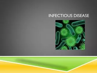
General Principles of Infectious Disease. November 2011 Lobna Al Juffali,MSc. Objectives. Recall foundational principles of microbiology, pharmacology, pathophysiology, & immunology in the treatment of infectious diseases

E N D
General Principles of Infectious Disease November 2011 Lobna Al Juffali,MSc
Objectives • Recall foundational principles of microbiology, pharmacology, pathophysiology, & immunology in the treatment of infectious diseases • Recognize predisposing conditions leading to the development of infection and preventative measures
Objectives Cont’d • Describe physical findings, lab etc used in the diagnosis of infection and monitoring of response to therapy • Name and differentiate the 3 primary uses of antibiotics • Explain the use of patient data to optimize initial and subsequent antibiotic therapy
Outline • Microbiology • Laboratory Tests to direct antimicrobial pharmacotherapy • Antibiotic Introduction • Systemic Approach in selecting an antibiotic
Pharmacotherapy of Infectious Diseases General Principles • Why do we care? • Major cause of morbidity & mortality • Accounts for billions $ a year world wide • Inpatient & Outpatient prescriptions • 1/3 of hospital budgets • 14 of the top 100 hospital drugs • Major % of outpatient prescriptions
Why does it happen? 1 We share the world with potential pathogens • Exposure to a virulent pathogen • Brucella, Malaria, HIV, Tb, STD’s, H1N1 • Public health measures are not followed like • Hand washing • Vaccination • Vector control • Avoiding contact
Factors predisposing to infection • Alteration in normal flora • Distruption of natural barriers • Age • Immunosuppression secondary to: • Malnutrition • Underlying disease • Hormones • drugs
Microorganisms • Gram-positive (Cocci, Bacilli) • Aerobic • Anaerobic • Gram-negitive (Cocci, Bacilli) • Aerobic • Anaerobic
Gram-positive cocci Staphylococcus aureus Staphylococcus epidermidis Streptococcus (groups A, B, C, G) Streptococcus bovis Streptococcus pneumoniae Streptococcus viridans group Enterococcus faecalis E. faecium Gram-positive Bacilli Cornyebacterium Listeria Bacillus Gram-negative cocci Moraxella (Branhamella) catarrhalis Neisseria gonorrhoeae Neisseria meningitidis Aerobic Microorganisms
Gram-negative bacilli Enterobacteriaceae Citrobacter Enterobacter Escherichia coli Klebsiella Proteus Salmonella Shigella Serratia marcescens Yersinia Providencia stuartii Acinetobacter Campylobacter Haemophilus influenzae Helicobacter pylori Pseudomonas aeruginosa Legionella spp Stenotrophomonas (Xanthomonas) maltophilia Aerobic Microorganisms
Gram-positive cocci Peptococcus Peptostreptococcus bacilli Clostridia C.perfringens C. difficile C. tetani C. botulinum Propionibacterium acnes Gram negitive Bacilli Bacteroides fragilis Prevotella Fusobacterium Anaerobic Microorganisms
Chlamydiae C. pneumoniae C. trachomatis Mycoplasmas M.pneumoniae Spirochetes Treponema pallidum Borrelia burgdorferi Rickettsiae Mycobacteria Mycobacterium tuberculosis Mycobacterium avium intracellulare Viruses (Hepatitis, Influenza, HIV) Fungus (Candida, Aspergillus) Protoza Miscellaneous microorganisms
Normal flora • The human body contains a vast variety of microorganisms that colonize body systems These organisms occur naturally in the tissues of the host and provide some benefits , including: • defense by occupying space • competing for essential nutrients with pathogenic bacteria • stimulating cross-protective antibodies • suppressing the growth of potentially pathogenic bacteria and fungi
The effect of Antibiotics on normal flora Pharynx • oral thrush Intestine • pseudomembranous colitis • Colonization with resistant organisms
Laboratory tests confirming the presence of infection • NONSPECIFIC TESTS • White blood cell count and differential • Other tests • the erythrocyte sedimentation rate (ESR) • the C-reactive protein concentration, • they are elevated in an inflammatory process but do not confirm the presence of infection because they are often elevated in noninfectious conditions, such as collagen-vascular diseases and arthritis. • Large elevations in ESR are associated with endocarditis, osteomyelitis, and intraabdominal infections. • TNF alpha found in patients with serious infections.
Laboratory Identification of pathogens • Direct examination (gram-positive, gram-negative, gram-variable, bacillus, or cocci). • Microscopic examination • Gram stain • Cultures
Microscopic examination wet-mount specimen preparations can provide valuable information regarding potential pathogens. Sputum bronchial aspirates scrapings of mucosal lesions urinary sediment.
Gram stain • Using crystal violet and iodine • Cerebrospinal fluid (CSF) in cases of suspected meningitis, • on urethral smears for venereal diseases, • on abscess or effusion specimens. • Sputum • They are helpful in identifying organisms that may not grow on culture and which otherwise would be missed.
Other staining Techniques • For Fungi • India ink • potassium hydroxide (KOH) • Giemsa stains • ForMycobacterierium tuberculosis or atypical mycobacteria • Ziehl-Neelsen stain for acid-fast bacilli
Cultures • Isolation of the etiologic agent by culture is the most definitive method available for the diagnosis and eventual treatment of infection. • Every effort should be made • to avoid contamination. • Time(perish from air or dry) • Transport media • Source of specimen should be recorded
Diagnosis of Infection using immunologic Methods(serology testing) • Antibody and antigen Detection • detected easily during acute infection • Immunofluorescence, • cytomegalovirus, respiratory syncytial virus, • Latex agglutination • meningococcal capsular antigens in CSF of patients suspected of having bacterial meningitis • Legionellapneumophila. • Enzyme-linked immunosorbent assay (ELISA) • HIV,
Susceptibility • The minimum inhibitory concentration MIC is defined as the lowest antimicrobial concentration that prevents visible growth of an organism after approximately 24 hours of incubation in a specified growth medium. • The minimum bactericidal concentration MBC is • is defined as the lowest concentration of drug that kills 99.9% of the total initially viable cells (representing a 3 log CFU/mL or greater reduction in the starting inoculum). • It is used in assessing the treatment of more severe infections, such as endocarditis and osteomyelitis
Susceptibility • Susceptible (S) • Clinical success can be expected if treated with usual doses • Intermediate (I) • Clinical success may be possible if • High doses of antibiotic are used • Antibiotic concentrates at the site of infection • Combination of synergistic agents are used • Resistant (R) • Treatment failure is expected
Quantitative antimicrobial susceptibility Testing • The minimal inhibitory concentration (MIC) • The MIC quantitatively determinates in vitro antibacterial activity. determined via • the macrotube dilution method • 96-well microtiter plates • Solid agar
Qualitative antimicrobial susceptibility Testing Disc diffusion assay
The Primary uses of antibiotics • Prophylaxis • Medical • Surgical • Empiric Treat likely / suspected pathogens (usually up to 72 hours) • Definitive Treat known / confirmed susceptible pathogen Use the most effective, least toxic, narrowest spectrum, and most cost effective agent (Drug of Choice)
Pharmacodynamic • Bacteriostatic • Inhibits growth at all concentrations above MIC • Requires intact immune system for killing • Avoid in life-threatening diseases states • Still may be a drug of choice if no other options
Pharmacodynamic Cont’d • Bacteriocidal: • Kills MO above MIC, Kills above MBC • Dose Dependent Killing (Peak to MIC) • Aminoglycosides and Quinolones • “once daily” aminoglycosides • Time Dependent Killing (Time Above MIC) • Beta-Lactams
POSTANTIBIOTIC EFFECT is the persistent suppression of an organism’s growth after a brief exposure to an antibiotic A PAE equal to or greater than 1 hour has been demonstrated for most antibiotics against gram-positive bacteria. As a general rule, antibiotics that inhibit DNA or protein synthesis (e.g., quinolones and aminoglycosides) demonstrate significant PAEs against gram-negative organisms.
POSTANTIBIOTIC EFFECT The primary clinical application of the PAE is to allow for less frequent administration of antimicrobials while still maintaining adequate antibacterial activity (e.g., extended-interval aminoglycoside administration).
Pharmacokinetics (ADME) • Absorption • Many antibiotics are IV only or PO only • Others have excellent oral bioavailability • safer / outpatient treatment • Distribution • Many sights of infection are not easily reached by antibiotics • Central nervous system, lung, bone
Pharmacokinetics (ADME) • Metabolism / Eliminations • Hepatic: drug interactions via CYP 450 • Inhibitors: Macrolides, Azoles • Inducers: Rifampin • Both: Protease inhibitors • Renal: dose adjustment with dysfunction • Elderly, critically ill
TIMING OF COLLECTION OF SERUM SAMPLES Peak and/or trough concentrations are monitored routinely for only a select few antimicrobials(e.g.,aminoglycosides and vancomycin) It is crucial to ensure that the antimicrobial’s administration time and serum sample time(s)are recorded Samples ideally should be obtained after steady state is achieved
Combination therapy Synergy Combination of the two antibiotic is Significantly greater than the sum of activity of either agent alone Antagonism combination may result in activity that is worse than either agent alone indifferent or additive. Combination activity that is neither synergistic nor antagonistic
Aminoglycosides • Serum conc. Has been linked to clinical response and nephrotoxicity • once-daily versus multiple daily aminoglycoside dosing
Vancomycin • vancomycin has been associated with oto- and nephrotoxicity in humans, • most of these reports occurred with • older, impure formulations of the drug • with extremely high concentrations uncommon with contemporary dosing regimens • or when vancomycin was combined with known nephrotoxic agents. • continuous infusions versus intermittent regimens
Systematic Approach for Selection of Antimicrobials • Confirm the presence of infection • Careful history and physical • Signs and symptoms • Predisposing factors • Identification of the pathogen • Collection of infected material • Stains • Serologies Culture and sensitivity • Selection of presumptive therapy considering every infected site • Host factors • Drug factors • Monitor therapeutic response • Clinical assessment • Laboratory tests • Assessment of therapeutic failure
CONFIRMING THE PRESENCE OF INFECTION • Fever • Normal body temperture 36.7 to 37 ˚C • Single oral temp >38.3˚ C or 38.0 ˚C over at least 1 hour • Rectal Temp - subtract 0.6˚ C (1 F) • Axillary - add 0.6˚ C (1 F) • Fever is a manifestation of many disease states other than infection. • Drug-induced fever is defined as persistent fever in the absence of infection or other underlying condition.
CONFIRMING THE PRESENCE OF INFECTION White Blood Cell Count (WBC) • The number of leukocytes (WBC) in the blood is often an indicator of disease.4000 and 10,000/mm3 • An increase in the number of leukocytes over the upper limits is called leukocytosis • A decrease below the lower limit is called is called leukopenia. • Most infections result leukocytosis because of the mobilization of granulocytes and/or lymphocytes to destroy invading microbes.