Download

1 / 62
661 likes | 1.12k Views
Thyroid Cancer. Megan Mezera, MD Resident, PGY-2 James Graham Brown Cancer Center Department of Radiation Oncology. Thyroid Overview. Thyroid Anatomy. Thyroid is a gland that consists of right and left lobes, joined by an isthmus . It extends from vertebral levels C5 to T1 .

E N D
Thyroid Cancer Megan Mezera, MD Resident, PGY-2 James Graham Brown Cancer Center Department of Radiation Oncology
Thyroid Anatomy • Thyroid is a gland that consists of right and left lobes, joined by an isthmus. • It extends from vertebral levels C5 to T1. • It lies anterior to the 2nd - 4th tracheal rings. • A pyramidal lobe may extend superiorly from the isthmus or from one of the lateral lobes. • Average weight is 20 g. • Parathyroid glands are located on the posterior surface of both thyroid lobes. • The recurrent laryngeal nerves lie in a cleft between the trachea and esophagus, medial to the lateral aspect of both thyroid lobes.
Anatomy cont. • Inner true capsule: Thin and adheres closely to the gland. • Extensions of this capsule within the gland divide it into lobes & lobules. • The lobules are composed of follicles, the structural units of the gland. • Follicles consist of a layer of simple epithelium enclosing a colloid-filled cavity. • Epithelial cells are of 2 types: • Principal cells (follicular cells). • Responsible for formation of colloid (Iodothyroglobulin). • Parafollicular cells (C cells, clear cells, light cells). • Produce the hormone Calcitonin, a protein central to calcium homeostasis.
Histology of thyroid gland: Follicles = structural units of the gland, consisting of a single layer of simple epithelium enclosing a colloid-filled cavity.
Lymphatic Drainage • Internal Jugular chain of Cervical Lymph Nodes • Superior thyroidal – levels 2, 3, 6 • Inferior thyroidal – levels 4, 6, paratracheal • Level VI: • Pre-laryngeal (Delphian) node • Recurrent laryngeal nodes • Pre- & Para-tracheal nodes • Anterior-superior mediastinal nodes are secondary nodes to the recurrent laryngeal and pre-tracheal nodal groups. Image: Japanese Journal of Clinical Oncology. Vol. 41, Issue 8.
Physiology and Function • Thyroid gland manufactures two essential hormones: • Thyroxine (T4). • Tri-iodothyronine (T3). • There is minimal difference between T3 and T4. The numbers refer to the amount of atoms of iodine contained in the hormones. • T3 is more potent, and is the more active form in the target tissues. • T4 is released by the thyroid in larger amounts, but is mostly converted to T3 in the liver and kidneys. • TSH from the pituitary regulates secretion of T3 & T4. • Effects of T3 and T4: • Increase the basal metabolic rate of most cells in the body. • Increase fat and carbohydrate metabolism. • Boost protein synthesis. • Increase heart rate and blood flow to other organs.
Epidemiology • Thyroid cancer is uncommon. • 1-2% of all malignancies. • < 0.2% of all cancer deaths in the United States. • Incidence is increasing. • However, may be due to increased detection of subclinical disease by extensive use of ultrasound, FNA. • American Cancer Society estimates: • 30,180 new thyroid cancer cases estimated in 2006 (7,590 men and 22,590 women). • 1,500deaths (630 men and 870 women) from the disease. • Male:Female = 1:3 • Incidence peaks in 5th decade. • Radiation exposure is main environmental risk factor.
Thyroid Cancer - types • Papillary – 75-80% • Follicular cell origin. • Tends to grow slowly. • Follicular – 15 % • Also begins in the follicular cells. • Medullary – 5-10% • Only thyroid cancer that originates from the Parafollicular C cells. • Anaplastic – 1-2% (rare) • Follicular cell origin. • Aggressive. Tends to grow and spread very quickly. Other rarer types (Non-Epithelial origin) – < 5% • Sarcomas • Malignant Lymphomas (NHL) • Hemangio-endotheliomas • Metastases from other primary sites • Thyroid cancer arising in unusual sites (ex: Struma Ovarii).
Papillary Thyroid Carcinoma (PTC) • Age 30-40 years • 1/3 have clinical LAD at presentation. • About 50% of excised nodes are positive. • Primary disease is confined to the neck in 95-99% cases at diagnosis. • Distant mets are rare (1-7%). • 75% are multi-focal. • May have a large follicular component. – Mixed Papillary/Follicular cancers • Tall cell and Insular carcinomas are variants with a worse prognosis
PTC – Pathology • Arise from follicular cells of the thyroid • Psammoma bodies - round collection of calcium • Orphan Annie Eye - empty or ground glass nuclei
Follicular Thyroid Carcinoma (FTC) • Usually age >50 years • Tends to be slightly more aggressive than PTC. • Average tumor size is larger than PTC. • Metastatic spread to other parts of the body is more common in FTC. • Most common sites of mets are lung and bones. • Rare clinical LAD at presentation (4-6%). • Thyroglobulin can be used as a tumor marker for well-differentiated FTC. • Hurthle cell and primary Clear cell carcinoma are variants.
FTC - Pathology • Cannot be diagnosed on FNA due to inability to distinguish between benign follicular adenoma and carcinoma. • Scalloping of follicles
Hürthle Cell Carcinoma • Thought to be a variant of Follicular Thyroid Ca. • Can be benign or malignant. • If no evidence of invasion, tend to act like adenomas. • If malignant, typically more aggressive than FTC or PTC. • 10-year survival 76% vs. 85% • Microscopically characterized by abundant eosinophilic granular cytoplasm.
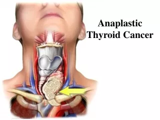
Anaplastic Thyroid Carcinoma (ATC) • Least common type (<5%). • Occurs late, >60 yrs age. • History of goiter in 80% of patients. • Possible transformation from benign to malignant lesion. • Highly malignant, with rapid invasion of the adjacent structures and metastases throughout the body. • Pathology may reveal evidence of PTC or FTC which may represent the precursor lesion. • Histology is atypical with Spindle-shaped cells and Multi- nucleated Giant cells • All pts with ATC are considered Stage IV. • Poor prognosis: Death usually occurs within 12 months of diagnosis.
Medullary Thyroid Carcinoma (MTC) • The only thyroid cancer that originates from the Para-follicular/C cells. • Two types of MTC: • Sporadic (80%) • Familial (20%) • Familial MTC: • Bilateral. • Less likely to have cervical nodes involved at diagnosis. • Better prognosis. • Patients with familial MTC should consider screening tests for genetic abnormalities – RET proto-oncogene (MEN syndromes). • Hypersecretion of Calcitoninused as a marker of disease status.
MTC - Pathology • May have considerable fibrosis • Amyloid deposits • Immunohistochemical staining with anti-calcitonin antibody
Multiple Endocrine Neoplasia (MEN) Syndromes • MEN-2a : (60 to 90% of MEN cases) • High lifetime risk of medullary thyroid cancer (~ 90%). • Average age of MTC diagnosis between 15 and 20 years. • 95% have a mutation in the RET proto-oncogene (Chromosome 10). • 95% have an affected parent, while 5% are de novo. • Pheochromocytoma & Parathyroid Adenoma. • MEN-2b (or 3) : (5% of MEN cases) • MTC. • Pheochromocytoma. • Marfanoidhabitus. • Mucosal neuromas of tongue and lips. • Neurofibromas. • FAMILIAL MTC: (5 to 35% of MEN cases) • No pheochromocytoma or parathyroid disease. • Mutation in RET proto-oncogene. • Age of onset of MTC later than with MEN 2A. PROPHYLACTIC THYROIDECTOMY HAS >90% CURE
Thyroid Cancer - Genetics • Papillary Thyroid Carcinoma • Somatic point mutations in the BRAF gene. • Medullary Thyroid Carcinoma • Gain of function mutation in RET proto-onco gene located on Chromosome 10.
Survival • Papillary and Follicular: 95-97% DSS at 20 yrs. • Hurthle Cell Ca: 80% DSS at 10 yrs. • Medullary Ca: 60-80% DSS at 10 yrs. • Anaplastic Ca: 25-45% DSS at 1 yr.
Radiation-Induced Thyroid Cancer • Exposure to ionizing radiation, particularly before puberty. • 25% of patients who receive as low as 0.02 Gy external irradiation to the thyroid gland develop goiters. • 25% of these, or 7%of all individuals who receive external irradiation to the thyroid, develop cancer. • Usually Papillary Ca. • In the past, external irradiation was used to treat children with conditions such as acne, fungal infections of the scalp, enlarged thymus gland, or to shrink enlarged tonsils or adenoids.
Radiation-InducedThyroid Cancer cont. • Children are much more sensitive than adults. • Histology: well-differentiated, develops slowly. • Mortality rates are low: <5% die due to RT-induced Ca. • Risk of cancer follows a linear dose response curve without threshold. • Even when the thyroid gland is outside of the field of radiation, an increased risk of thyroid cancer has been noted following radiation treatment in childhood for Hodgkin and non-Hodgkin lymphomas, ALL, Wilm's tumor, neuroblastoma, and central nervous system tumors (Inskip PD, Med PediatrOncol. 2001). • No increased risk for cancer is seen after diagnostic I131.
History • External irradiation to head, neck, or chest between infancy and early adulthood. • Family history of medullary thyroid cancer • Inherited as an Autosomal Dominant condition. • Family or personal history of pheochromocytoma or hyperparathyroidism with or without mucosal neuromas • Suggestive of MEN syndromes. • Most cases are spontaneous.
Clinical Manifestations & Presentation • Painless lump in the neck. • Representing either primary thyroid tumor or regional lymph nodes. • Rapid growth in suspected benign thyroid nodule. • Clinically occult in some cases (seen on imaging only). • Pts with thyroid CA are typically EUTHYROID. • Normal TSH levels. • Voice change, hoarseness. • Suspect recurrent laryngeal nerve involvement or impingement. • Dysphagia • Difficulty breathing
Prognostic Factors • Age is most important factor. • Increased mortality in patients >45 years old • Tumor size >4cm • Higher histologic grade • Hurthle cell variety • Post-op macroscopic residual disease • Male sex • Presence of distant mets - Rao RS, et al. Prognostic factors in follicular carcinoma of the thyroid: a study of 198 cases. Head Neck 1996;18:118–126 - Shaha AR, et al. Prognostic factors and risk group analysis in follicular carcinoma of the thyroid. Surgery 1995;118:1131–1136.
T- Staging (AJCC 2010) • Papillary/Follicular/Medullary Carcinomas: • T1: Tumor ≤2 cm, Intra-thyroidal • T2: Tumor >2 cm but not >4 cm, Intra-thyroidal • T3: Tumor >4 cm in greatest dimension limited to the thyroid, or any tumor with minimal extrathyroid extension (e.g., extension to sternothyroid or perithyroid soft tissues) • T4a: Tumor of any size extending beyond the thyroid capsule to invade subcutaneous soft tissues, larynx, trachea, esophagus, or recurrent laryngeal nerve • T4b: Tumor invades prevertebral fascia or encases carotid artery or mediastinal vessels. • All anaplastic carcinomas are considered stage IV tumors. • T4a: Intrathyroidalanaplastic carcinoma — surgically resectable • T4b: Extrathyroidalanaplastic carcinoma — unresectable
N & M Staging • N0: No regional lymph-node metastasis • N1: Regional lymph-node metastasis • N1a: Metastasis to level VI • Pretracheal, paratracheal, and prelaryngeal/Delphian lymph nodes • N1b: Metastasis to unilateral, bilateral, or contralateral cervical or superior mediastinal lymph nodes • M0: No distant metastasis • M1: Distant metastasis
Evaluation of Thyroid Nodule • Ultrasound of thyroid with FNA. • Sensitivity 98%, Specificity 99%, Accuracy 98% • Remember: Negative FNA does not exclude FTC. • Labs: T3, T4, TSH, Thyroglobulin • Medullary Ca: • Calcitonin, CEA, calcium • Urine and serum catecholamines (screen for pheochromocytoma) • Patient and family members should be screened for RET proto-oncogene • Ultrasound, MRI, non-contrast CT of neck to evaluate adenopathy • Do not use iodinated CT contrast as this will delay 131I treatment up to 6 months
Surgery • Total thyroidectomy favored over subtotal thyroidectomy • Recurrence rate (Mazzaferri, Am J Med) • Total thyroidectomy: 7% • Subtotal thyroidectomy: 18% • Total thyroidectomy leaves a small amount of residual thyroid tissue in order to avoid morbidity from damaging adjacent structures (i.e. recurrent laryngeal nerve) • Modified radical neck dissection or limited neck dissection favored over radical neck dissection • Lower morbidity with limited dissection • No survival benefit with more aggressive dissection
Post-Operative Therapy • Suppression of residual thyroid tissue with exogenous thyroid hormone (i.e. Levothyroxine) • Ablation of thyroid remnant with radioactive 131I • Addition of post-op thyroid hormone replacement or 131I ablation decreases recurrence rates vs. surgery alone. • Total thyroidectomy alone: 7% • TT + Thyroid Hormone or I-131: 2.6% • Thyroid Hormone alone: 10%
Post-Operative Therapy cont. • 214 patients with Follicular Ca (Young, J Nucl Med) • 10 year follow-up recurrence rates • Total thyroidectomy alone: 33% • TT + Thyroid Hormone: 10% • TT + TH + I-131: 6% • Only patient deaths due to disease had metastases at presentation
Radioactive 131I (RAI) • Taken up by follicular cells • Can be used to treat papillary, follicular (including Hürthle subtype), and some medullary cancers • C-cells do not take up RAI, but may consider for inoperable MTC as nearby follicular cells will take up RAI and may kill adjacent MTC cells in thyroid • Will not treat MTC-involved LN or distant mets
Indications for RAI • Tumor size >1.0 cm • Thyroid capsule invasion • Vascular invasion • Multifocal disease • Soft tissue invasion • Post-operative residual disease (+ margins) • Cervical or mediastinal nodal metastases (N1b) • Distant metastases • Recurrent disease • Other • Eliminates any residual normal thyroid tissue • Improves reliability of future iodine uptake scans and thyroglobulin levels for disease monitoring
Induction of 131I Uptake • Must make the residual thyroid tissue “hungry” for iodine • Two ways to encourage uptake of 131I by residual thyroid tissue • Make the patient hypothyroid • Low TH levels increases TSH promotes 131I uptake • Goal TSH >30 • Give recombinant TSH (rhTSH, Thyrogen) • Exogenous TSH promotes 131I uptake • Tolerated better by patients as does not induce hypothyroidism • Patients must remain on low-iodine diet prior to treatment
RAI Ablation • 131I is used • Physical t½ = 8 days • Radiation Dose: Total Body:Thyroid = 1:300-1000 • 100-200 mCi after preparatory regimen with a target TSH >30 or with Thyrogen (rhTSH). • Start/restart thyroid hormone replacement (Synthroid) • Diagnostic uptake scan 7-10 days after 131I • Demonstrates amount and location of thyroid tissue • If +ve,then re-scan 4-6 months later and may re-treat if persistently positive. • If –ve, then follow clinically with exam and thyroglobulin levels (though some MD’s re-scan in 1 year) • Never use iodinated contrast 3-6 months prior to RAI therapy.
Complications of RAI ablation • Acute: • Sialadenitis, xerostomia, cystitis, radiation gastritis, transient leukopenia and thrombocytopenia, transient oligo-spermia in males, thyrotoxicosis during the 1st two weeks from increased tumor lysis, radiation pneumonitis if pulmonary metastasis present. • Chronic: • Risk of leukemia with cumulative doses >800 mCi • Breast and bladder Ca with >1000 mCi • Bone and soft tissue Ca with >800-1000 mCi • Pulmonary fibrosis in patients with diffuse pulm mets - Robino C, Br J Cancer, 2003 • No increase in infertility, miscarriages, prematurity, or congenital anomalies. However, recommend that pts wait 6 months prior to attempting pregnancy.
Indications for EBRT • Primary therapy for locally unresectable thyroid cancer • Particularly if 131I does not concentrate in tumor • Bulky tumor (e.g. mediastinal disease) large enough that it is uncontrollable by 131I alone • Malignant cervical adenopathy that may not be controlled by 131I alone • Superior vena cava syndrome • Repeatedly recurring thyroid cancer regardless of 131I accumulation • Anaplastic Ca or Medullary Ca with high risk features
Post-Therapy Follow-up Tests • Thyroid function panel • Well-Differentiated CA (Papillary/Follicular CA) • Thyroglobulin level or ThyroglobulinAb level • Radioactive Iodine Uptake Scan • Make the patient hypothyroid to increase TSH • Give low activity radioactive iodine (123I or 131I) • Scintigraphy detects uptake of iodine and indicates residual thyroid tissue • Medullary CA • Calcitonin level • C Cells do not uptake iodine so RAI uptake scan is not useful
Significance of Thyroglobulin • Thyroglobulin is produced by the thyroid only • Glycoprotein function in the iodination of thyroid hormone • Pre-op value elevated: • Differentiated thyroid cancer • Follicular adenoma • Other benign thyroid diseases. • An elevated thyroglobulin level cannot differentiate benign from malignant lesions pre-operatively • Athyrotic patients should not have circulating thyroglobulin (t½=65 hr) • Post-op value elevated: • Indicates residual, recurrent, or metastatic differentiated thyroid cancer. • Correlates well with 131I imaging for detection of thyroid cancer.
Correlating 131I Scan and Thyroglobulin Levels • In 80% to 85% of patients, the thyroglobulin levels and 123I imaging results agree, with abnormal 123I scans seen in patients with elevated thyroglobulin levels. • In 15% to 20% of cases, the results do not agree. • Pineda et al, reported on patients with elevated thyroglobulin and negative diagnostic 131I scans. • They treated 17 patients with 131I fitting this scenario. • In half of their patients, the serum thyroglobulin level normalized after treatment . • Elevated thyroglobulin levels with normal 131I scan results is believed to reflect occult disease that is not detectable on imaging • Normal serum thyroglobulin levels occur in conjunction with an abnormal 131I scan in 1.5% of patients and probably reflect some decrease in tumor function or tumor differentiation.
Management – Papillary/Follicular/Hürthle • Low Risk Disease: • Age 15-45 • No distant mets • No extra-thyroidal extension • Tumor size <4 cm • Treatment: • Total Thyroidectomy • Thyroid hormone replacement with good TSH suppression • +/- radioactive iodine ablation
Management – Papillary/Follicular/Hürthle cont. • High Risk Disease: • Age <15 or >45 • Known regional or distant mets • Extra-capsular extension • Tumor size >4 cm • Treatment: • Total Thyroidectomy with LN sampling • LN dissection if positive nodes • Radioactive Iodine Ablation • If LN+, then consider EBRT following RAI. • Thyroid hormone replacement • Minimal role for chemo
Management – Medullary Thyroid Carcinoma • Total Thyroidectomy with lymph node dissection • EBRT indications: • Residual disease • Extensive nodal involvement • T4 • Neighbor effect role for RAI • Parafollicular (C) Cells do not uptake iodine, but adjacent follicular cells do and may concentrate adequate dose in the area • Thyroid hormone suppression has no effect because C Cells are not regulated by TSH • Minimal role for chemo
Management – Anaplastic Carcinoma • Complete surgical resection gives the only chance of cure. • If gross total resection not possible, then radical surgery is not indicated except for airway management. • EBRT +/- chemo used for local control and palliation. • Chemo – Doxorubicin based • EBRT – twice daily • No role for RAI ablation. • Predictors of better outcomes • Age <60 • Intra-thyroidal confined tumor • Combined use of surgery and EBRT
Tubiana M, et al. External Radiotherapy in Thyroid Cancers. Cancer, 1985 • Advocated post-operative irradiation for patients with residual disease. • 50 Gy in 25 fractions over 5 weeks to the neck with a boost of 5 to 10 Gy to residual disease with 60Co teletherapy at the Institut Gustave-Roussy, Villejuif, France • 5-year survival rate • 94% (62 of 66) for patients with complete surgery • 78% (76 of 97) for patients with incomplete surgery
Farahati J. Differentiated Thyroid Cancer. Cancer, 1996. • 238 patients with well differentiated thyroid carcinoma • 99 patients received adjuvant EBRT • Recurrence-free survival was improved with EBRT in patients • >40 years of age • LN+