
Introduction
Introduction. Many studies require the quantitative determination of bacterial populations. The two most widely used methods for determining bacterial numbers are: The standard plate count method. Spectrophotometer (turbid metric) analysis.

Introduction
E N D
Presentation Transcript
Introduction • Many studies require the quantitative determination of bacterial populations. The two most widely used methods for determining bacterial numbers are: • The standard plate count method. • Spectrophotometer (turbid metric) analysis. • The standard plate count method is an indirect measurement of cell density ( live bacteria). • The spectrophotometer analysis is based on turbidity and indirectly measures all bacteria (cell biomass), dead and alive.
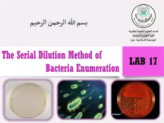
The Plate Count (Viable Count) • However, if the sample is serially diluted and then plated out on an agar surface in such a manner that single isolated bacteria form visible isolated colonies, the number of colonies can be used as a measure of the number of viable (living) cells in that known dilution. • The number of bacteria in a given sample is usually too great to be counted directly.
In addition, some of the bacteria may be clumped together. Therefore, when doing the plate count technique, we generally say we are determining the number of Colony-Forming Units (CFUs) in that known dilution. • By extrapolation, this number can in turn be used to calculate the number of CFUs in the original sample. • bacterial counts by these methods are usually expressed as colony forming units per milliliter (CFU/mL). • Keep in mind that if the organism normally forms multiple cell arrangements, such as chains, the colony-forming unit may consist of a chain of bacteria rather than a single bacterium.
A plate having 30-300 colonies is chosen because this range is considered statistically significant. • Normally, the bacterial sample is diluted by factors of 10 and plated on agar. • After incubation, the number of colonies on a dilution plate showing between 30 and 300 colonies is determined.
Likewise, if there are more than 300 colonies on the plate, there will be poor isolation and colonies will have grown together. (too numerous to count (TNTC). • If there are less than 30 colonies on the plate, small errors in dilution technique or the presence of a few contaminants will have a drastic effect on the final count. (too few to count (TFTC).
Procedure • Using sterile technique, transfer 1 mL sample to the first dilution blank. Mix the bottle by inverting it 20 times. Label the bottle "10-1."
Using a fresh pipette, transfer 1 mL from the first blank to the second blank. Mix as before. Label the second bottle "10-2“. • Using a fresh pipette, transfer 1 mL from the first blank to the third blank. Mix as before. Label the second bottle "10-3“. • Using a fresh pipette, transfer 1 mL from the first blank to the forth blank. Mix as before. Label the second bottle "10-4“. • Using a fresh pipette, transfer 1 mL from the first blank to the second blank. Mix as before. Label the fifth bottle "10-5“. • Label the Petri dishes: 10-2, 10-3, 10-4, 10-5, and 10-6, respectively. Using a fresh pipette, transfer 1 mL from the first blank to the second blank. Mix as before. Label the second bottle "10-2."
One at a time, add a tube of molten nutrient agar to each Petri dish. After adding the agar, gently swirl the dishes in pattern for 30 seconds to mix the bacteria with the agar. After the agar has thoroughly solidified, incubate the plates at 37°C for 24 to 48 hours. Count the number of colonies on a plate that has between 30 and 200 colonies. Any plate which has more than 200 colonies is designated as "too numerous to count" (TNTC). Plates with fewer than 30 colonies do not have enough individuals to be statistically acceptable. transfer liquid from the dilution blanks to the Petri dishes. Use a separate pipette for each blank, not for each plate (i.e. if more than one plate uses liquid from a single blank, a single pipette may be used for that blank).
Colonies Forming Units {CFU} • Calculate the number of bacteria (CFU) per milliliter or gram of sample by dividing the number of colonies by the dilution factor multiplied by the amount of specimen added to agar plate. • To compute the number of CFU/mL, use the formula: • c = concentration, CFU/mL • n = number of colonies • d = dilution blank • s = volume transferred to plate.
CFU Calculation Example • You count 46 colonies on your plate • You put 1 ml of bacterial culture into 99 ml of saline and plated 0.1 ml • Dilution 1/100 CFU= 46 1/100 * 0.1 = 46 * 100 * 10 =46000 CFU/ml


