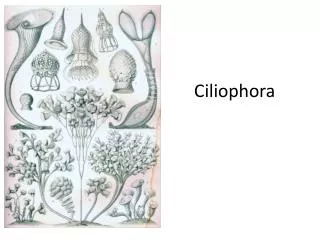
Chapter 10 - Phylum Ciliophora
Chapter 10 - Phylum Ciliophora. Taxonomy Phylum Ciliophora Class Litostomatea Order Vestibuliferida Genus Balantidium Class Oligohymenophorea Order Hymenostomatida Genus Ichthyophthirius. Phylum Ciliophora

Chapter 10 - Phylum Ciliophora
E N D
Presentation Transcript
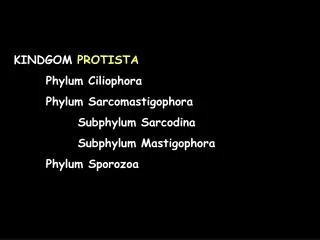
Chapter 10 - Phylum Ciliophora Taxonomy Phylum Ciliophora Class Litostomatea Order Vestibuliferida Genus Balantidium Class Oligohymenophorea Order Hymenostomatida Genus Ichthyophthirius
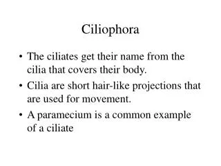
Phylum Ciliophora • Possess cilia simple cilia or compound ciliary organelles during some part of their life cycle • Most species have 2 kinds of nuclei: macronuclei and micronuclei • Some members of the phylum engage in sexual reproduction, involving conjugation, autogamy, and ctyogamy • Most ciliates are free-living; however, a few groups are commensals or parasitic • Important taxonomic criteria for members of this group include: structure of the cortex and arrangement of kinetosomes
Order Vestibuliferida • Members of the Order Vestibuliferida typically have cilia uniformly distributed over the body • All members of the order have a densely ciliated vestibulum near the apex of the cell • Vestibulum(peristome) is a depression or invaginated area that leads directly to the cytosome; it is lined with cilia
Family Balantidiidae • Family Balantidiidae, which includes only one genus and species (Balantidium coli)are found in the intestinal tract of arthropods and some vertebrates, including mammals • Pathogens of humans, pigs and monkeys
Balantidium coli • Morphology • Conspicuous vestibulum leads into a large cytostome; opposite of which lies a cytopyge • Coarse cilia line the peristomal area • Macronucleus is typically elongate and kidney-shaped; micronucleus is spherical • 2 prominent contractile vacuoles, indicating osmoregulation • Food vacuoles in the cytoplasm contain debris, bacteria, RBCs, and fragments of host epithelium
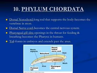
Life Cycle • Both a motile trophozoite and a cyst stage occur • The trophozoite inhabits the cecum and colon of humans and is the largest known protozoan parasite of humans • The cyst wall is very thick and possibly consists of 2 membranes • Transmission from one host to another is accomplished via the cyst • Encystation usually occurs in the large intestine but may also occur outside the body of the host • Cysts are common in the feces of the infected host • Infection occurs when contaminated food or water is ingested • Excystation occurs in the small intestine
Epidemology • Balantidiosis is most often found in tropical regions throughout the world • It is not a common human disease; the infection rate is less than 1% • The parasite is nonpathogenic in pigs and is much more prevalent (20-100%) among these hosts • Pigs are a good source of infection for humans in areas where they share habitation
Symptomatology • Trophozoites primarily resides in the cecal area and throughout the large intestine • Also thrive in the small intestine, an area rich in starch, but do not invade the intestinal mucosa • Proclivity for starch may be the reason for the trophozoite’s invasive character once it becomes established in the human cecal region, a region low in starch content • Interestingly, in the pig’s intestine, where starch is more abundant, the organism remains in the lumen • Trophozoites presumably secrete proteolytic enzymes that act upon the mucosal epithelium, facilitating tissue invasion • Results from infection range from asymptomatic to severe • Parasitic invasion of the mucosal lining is followed by hemorrhaging and ulceration - balantidine dysentery
Diagnosis • Examination of stool samples, looking for trophozoites and cysts • Trophozoites are readily identified because of their large size and the fact that B. coli is the only ciliate that parasitizes humans • The infection may disappear spontaneously or the host may become asymptomatic, with the host remaining as a carrier • Several drugs that are taken orally are known to eliminate the infection
Order Hymenostamatida • Buccal cavity has a well defined oral ciliary apparatus; buccal cavity on the ventral surface • Members of this group are heavily ciliated; cilia uniform • Family Ichthyophthiriidae • The family has one genus that contains two species; we will discuss only one, Ichthyophthirius multifiliis • I. multifiliis is a relatively common parasite of FW aquarium fish and fish farms
Ichthyophthirius multifiliis • Morphology • Large horseshoe shaped macronucleus that encircles a smaller micronucleus • May have several contractile vacuoles • Cytopyge found at the posterior end
Life Cycle • Attacks the epidermis, cornea and gill filaments of FW fishes • The mature trophozoites form pustules in the skin of their hosts • When the pustules rupture, they are released and usually settle on vegetation or the bottom sediments Trophozoite under the epidermis
Life cycle cont. • Each trophozoite t then forms a gelatinous cyst and undergoes asexual reproduction producing up to 1000 infective cells • The daughter trophozoites (tomites) represent the infective stage of the life cycle • It uses a long filament that emerges from a conical depression in the pellicle to burrow into the host’s skin • At this time it becomes a trophozoite and begins to ingest host tissue; pustules form to complete the life cycle
Treatment of aquarium fish can involve dilute concentrations of formaldehyde or methylene blue