SLE,SCLERODERMAMCTD
SLE,SCLERODERMAMCTD. Prof.Abdurhman Saud Alarfaj BSc.,MB.,CHB.,FRCP ( uk ).,FRCP(C).,FACP.,FACR. Systemic lupus erythematosus (SLE). Systemic lupus erythematosus (SLE) Definition

SLE,SCLERODERMAMCTD
E N D
Presentation Transcript
SLE,SCLERODERMAMCTD Prof.Abdurhman Saud Alarfaj BSc.,MB.,CHB.,FRCP(uk).,FRCP(C).,FACP.,FACR
Systemic lupus erythematosus (SLE) Definition • chronic, multisystem inflammatory disease characterized by autoantibodies directed against self-antigens, immune complex formation, and immune dysregulation resulting in damage to essentially any organ. Background: • First written description in13th century( Rogerius) named it lupus( Latin for wolf) as cutaneous similar to a wolf bite. • Osler recognized systemic features without skin . • Diagnosis with (LE) cells in 1948. • In 1959, anti-DNA.
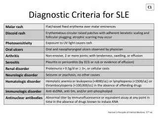
Hochberg MC. Updating the American College of Rheumatology revised criteria for the classification of systemic lupus erythematosus [letter]. Arthritis Rheum 1997;40:1725.
EPIDEMIOLOGY: • Locally: • 2 cases of SLE among 10,372 studied (prevalence of 19.28 per 100,000). • Internationally: variable prevalence :. • Denmark (21.7/100,000). • Britain, 12 cases per 100,000. • India prevalence (3.2/100,000) . • 39 cases per 100,000 population in Sweden.
AETIOLOGY: • Specific cause(s) of SLE is unknown. • multiple factors are associated include : • Genetic • Hormonal • Racial • Environmental factors
AETIOLOGY(cont.): • Genetic predisposition : • Multitude of genetic associations suggests a complex genetic predisposition. • Concordance rate in monozygotic twins is 25-70%. • If a mother has SLE, her daughter's risk of developing the disease is 1:40, and her son's risk is 1:250. • Relatives have a high prevalence of other autoimmune diseases. • HLA-DR2 and HLA-DR3 and other HLA genes occur more often in SLE than in the general population. • null complement alleles and congenital deficiencies of complement ( C4, C2, and other early components) are associated with an increased risk of SLE.
AETIOLOGY(cont.): • Hormonal factors: • F:M ratio of prevalence in different age groups: • In children, f:m ratio is 3:1 . • In adults, f:m ratio is 10-15:1 • In older, the ratio is approximately 8:1 . • Age at onset : • 65% have onset between 16 and 55. • 20% before age 16 , and • 15%t after age 55 . • Higher prevalence in men with Klinefelter disease. • Exogenous estrogen and exacerbations of SLE. • Men at all ages have the same risk of disease as women who are prepubertal or postmenopausal • Males do not have an age-related peak in incidence.
AETIOLOGY(cont.): • Racial andgeography : • Higher prevalence (2.5- to 6-fold) in USA African American women than in white women. • But,cf occurs infrequently in Blacks in Africa . • Higher among Asians, Afro-Americans, Afro-Caribbeans, Hispanic Americans, and Asian Indians. • More common in urban than rural areas . • Also In New Zealand, 50 per 100,000 Polynesians, but only 14.6 cases per 100,000 in the whites. • In France, more common among immigrants from Spain, Portugal, North Africa, and Italy .
AETIOLOGY(cont.): • Environmental: • worldwide variability of prevalence the disease(black in africa and US) • influence of environmental factors on the course of the disease, eg: • ultraviolet light • viruses • drugs.cause or exacerbate • silica dust. • cigarette smoking. • alfaalfa sprouts.
Pathophysiology: • Disturbances in the immune system : • High ratio of CD4+ to CD8+ T cells. • Defects in immune cell tolerance leading to • production of autoantibodies targeting antigens located in nuclei, cytoplasm, on cell surfaces, and in plasma proteins. • autoantibodies leads to mostly immune complex formation (e.g kidney) and direct antibody-mediated cytotoxicity (hemolytic anemia, thrombocytopenia). • Cell-mediated autoimmunity also play part. • Tissue damage follows
ORGAN INVOLVEMENT IN SLE Joints 90% Skin -Rashes 70% -Discoid lesions 30% -Alopecia 40% Pleuropericardium 60% Kidney 50% Raynaud’s 20% Mucous membranes 15% CNS (psychosis/convulsions) 15%
SLE – Presenting and Prevalent Symptoms ARA Criteria [n = 624] SAUDI ARABIA *In addition to those +ve at presentation
Primary Central Nervous System Lupus: Neurologic Signs or Symptoms Meninges Cerebellum Headache Ataxia MeningismusSpine Cerebrum Paraparesis Dementia Multiple sclerosis-like disorder Strokes Cranial and peripheral nerves Subarachnoid hemorrhage Cranial and peripheral sensory,motor neuropathies Migraine Mononeuritis multiplex Other headaches Myasthenia gravis Seizures Guillain-Barre syndrome Chorea Rigidity, tremor SIADH
Special considerations: • Drug-induced lupus(consider before diagnosing native lupus) • Sex ratios are nearly equal. • Nephritis and CNS not common. • No anti- native DNA or hypocomplementemia. • resolution on discontinuation of drug.
Drugs associated with lupus erythematosus • Possible Association • Betablockers • Methimazole • Captopril • Nitrofurantoin • Carbamazepine • Penicillamine • Cimetidine • Phenytoin • Ethosuximide • Propylthiouracil • Hydrazines • Sulfasalazine • Levodopa • Sulfonamides • Lithium • Trimethadione • Unlikely Association: • Allopurinol, • Penicillin, Chlorthalidone, Phenylbutazone, Gold salts, Reserpine,Griseofulvin,Streptomycin,Methysergide,Tetracyclines,Oral contraceptives • Definite association • Chlorpromazine • Methyldopa • Hydralazine • Procainamide • Isoniazid • Quinidine
TREATMENT (cont.): • GENERAL CONSIDERATIONS : • Prevention: • Avoid uv light and sun (sunsceening). • Antimalarial to prevent relapses. • Treat hypertension and dyslipidemias . • Treat depending on the organ system(s) involved: • Skin, musculoskeletal, and serositis. • NSAIDs,HCC,local cs. • More serious organ involvement( CNS,renal ) • Immunosuppression with high-dose steroids,AZA and/or cyclophosphamide,mycophenolate ,Tacrolimus • Targeted therapy(biological) ,rituximab • Other treatments • plasma exchange for TTP or diffuse alveolar hemorrhage • and intravenous immunoglobulin for severe steroid-nonresponsive thrombocytopenia.
PROGNOSIS : • Poor prognostic factors for survival in SLE include : • Renal disease (especially diffuse proliferative glomerulonephritis). • Hypertension • renal and central nervous system (CNS) disease • less education (?poor compliance) • Poor socioeconomic status (?inadequate access to medical care ). • Black race (? low socioeconomic status) • Presence of antiphospholipid antibodies • High overall disease activity • Male sex • Men similar freq of renal,skin,arthritis,and CNS as women, • but less photosensitivity, • more serositis, • an older age at diagnosis, • and a higher one year mortality. • Young age • SLE in children more severe,higher malar rashes, nephritis, pericarditis, hepatosplenomegaly, and hematologic abnormalities .
Remission – • After appropriate therapy, • many patients go into a clinical remission requiring no treatment. • a long-term follow-up of 667 patients noted: • ≈25 % had at least one treatment-free clinical remission lasting for at least one year. • The mean duration of remission was 4.6 years ( ?underestimate since one-half of the patients were still in remission at the end of follow-up). • A long history of SLE or the presence of renal or neuropsychiatric disease did not preclude remission
CLASSIFICATION OF SCLERODERMA 1. Localized: Morphea: plaque like, guttate, generalized linear scleroderma Scleroderma ‘en coup de sabre’ (± facial hemiatrophy) 2. Generalized: With diffuse visceral involvement CREST syndrome Overlap with other connective tissue disease. 3. Chemical-induced scleroderma-like conditions e.g: vinyl chloride disease 4. Diseases with skin changes mimicking scleroderma e.g.: scleredema 5. Eosinophilic fasciitis
Progressive Systemic Sclerosis: Preliminary Diagnostic Criteria Patient must have major criterion or 2 minor criteria. Major criterion Proximal scleroderma Minor criteria Sclerodactyly Digital pitting or scars or loss of substance from finger pads Bibasilar paulmonary fibrosis
SYSTEMIC MANIFESTATIONS OF SCLERO-DERMA Pulmonary Gastrointestinal Renal Dyspnea Dysphagia Proteinuria Cough Dyspepsia Azotemia Hemoptysis Constipation Hypertension Pleuritic pain Diarrhea Renal failure Clubbing of nails Malabsorption Musculoskeletal Cardiovascular Polyarthralgia Arrhythmias Swelling of joints Myocardial failure Contractures
CLINICAL AND LABORATORY FEATURES OF MCTD ● Polyarthritis ● Raynaud’s phenomenon ● Swollen hands or sclerodactyly ● Abnormal esophageal motility ● Myositis ● Low incidence of lupus nephritis ● Hyperglobulinemia ● Positive ANA (often speckled pattern) ● Antibody to nRNP
Antibodies Associated with Rheumatic Diseases: Percentages of Patients Affected Antibodies to….. Percentages of patients Native DNA SLE: 50% - 60% Sm antigen SLE: 30% Histones Drug-induced SLE: 95% SLE: ≤ 60% Rheumatoid arthritis: 20% SS-A Sjogren’s syndrome: 70% SLE: 30% - 40% Scleroderma and mixed connective tissue disease: frequency and titers low SS-B Sjogren’s syndrome: 60% SLE: 15%
Antibodies Associated with Rheumatic Diseases: (continued) Antibodies to… Percentages of patients RNP Mixed connective tissue disease: 95% - 100% SLE: 30% at low titers Scleroderma: 10% - 20% Scl-70 Scleroderma: 10% - 20% Nucleolar antigens Scleroderma: 40% - 50% Centromere antigens CREST: 80% - 90% PM-1 Polymyositis: 50% Dermatomyositis: 10%