ENDOCRINE SYSTEM
Explore the functions of endocrine glands and hormones in maintaining body equilibrium, metabolism, and growth. Learn about the pituitary gland and its crucial role in hormonal regulation.

ENDOCRINE SYSTEM
E N D
Presentation Transcript
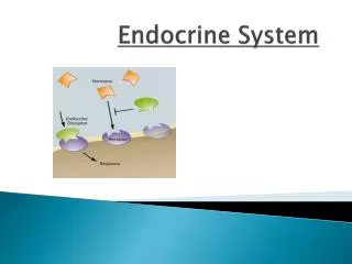
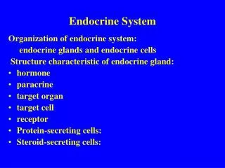
Introduction • The endocrine system consists of glands and tissues collectively called Endocrine Glands that secrete substances called hormones into the internal environment. • Endocrine glands are ductless; the hormones are secreted directly into the bloodstream for distribution throughout the body.
A HORMONE is a regulatory molecule that affects the activity of other glands or tissues. Hormones influence the metabolism of cells, the growth and development of body parts and homeostasis. • Most hormones are either steroids or steroid-like substances synthesized from cholesterol, amines, peptides, proteins or glycoproteins synthesized from amino acids. They can stimulate changes in the target cells even in extremely low concentrations.
GENERAL FUNCTIONS OF THE ENDOCRINE SYSTEM Regulating the functions of the human body to maintain homeostasis (equilibrium) is an enormous job. Two organ systems function coordinately to enable body parts to communicate with each other to adjust constantly to changing incoming signals. These are the nervous system and the endocrine system.
Endocrine glands and their hormones help regulate metabolic processes. They control the rate of certain chemical reactions and in the transportation of substances across membranes and help regulate water and electrolyte balance. • They also play a vital role in reproduction, development and growth
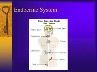
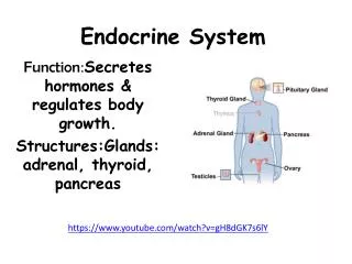
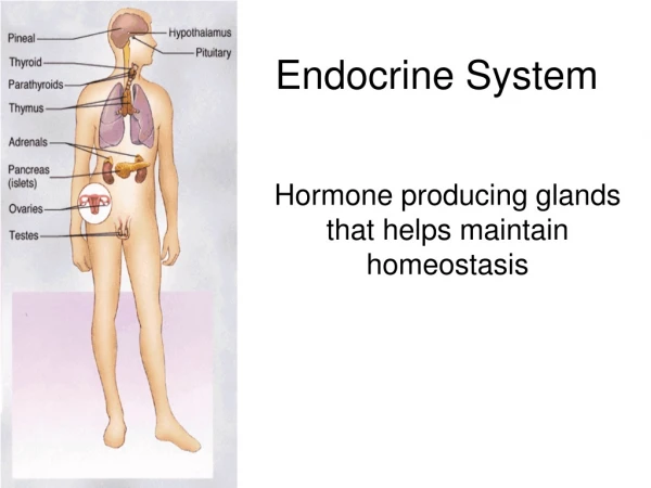
ENDOCRINE GLANDS Specialized small group of cells produce some hormones. However, the major endocrine glands are: • Pituitary gland • Thyroid gland • Parathyroid glands • Adrenal glands • Pancreas (Islets of langerhans) • Pineal gland • Thymus gland • Reproductive glands (testes and ovaries)
Although the hypothalamus is classified as a part of the brain and not as a direct endocrine gland it has a direct controlling effect on the pituitary and indirect effect on many other organs.
HYPOTHALAMUS AND PITUITARY GLAND • The hypothalamus and the pituitary glands act as a unit, regulating the activity of most of the other endocrine glands. • The pituitary gland (hypophysis) is located on the inferior (base) aspect of the brain where it is attached to the hypothalamus by a stalk- like structure called Infundibulum.
Roughly the size of a pea and about 1cm in diameter, the pituitary gland is known as the “master gland”. It weighs about 4g. The gland lies in the hypophyseal fossa of the sphenoid bone. • The pituitary gland is structurally and functionally divided into • anterior lobe (adenohypophysis) • posterior lobe (neurohypophysis). • These two parts have different embryonic origin.
The adenohypophysis is derived from a pouch of epithelial tissue that migrates upward from the embryonic mouth (pharynx) where as the neurohypophysis is formed as a down growth of nervous tissue of the brain.
In the fetus, a narrow region develops between the anterior and posterior lobes of the gland called the intermediate lobe (pars intermediate) which produces melanocyte –stimulating hormone (MSH), which regulates the synthesis of melanin – the pigment in the skin and in portions of the eyes and brain. • The intermediate lobe atrophies during prenatal development and in adults they no longer constitute a separate lobe or structure.
The posterior pituitary stores and releases hormones that are actually produced by the hypothalamus, whereas the anterior pituitary produces and secretes its own hormones. • The anterior pituitary however, is regulated by hormones secreted by the hypothalamus, as well as feedback from the target gland hormones.
Blood supply • Arterial blood is supplied by branches from the internal carotid artery. The anterior lobe is supplied indirectly by blood that has already passed through a capillary bed in the hypothalamus. • This network of blood vessel, the pituitary portal system, conveys blood from the hypothalamus to the anterior lobe. • As well as conveying oxygen and nutrients, this blood conveys the releasing and inhibiting hormones secreted by the hypothalamus that influence the secretion and release of hormones formed in the anterior lobe.
The posterior lobe is supplied directly by a branch from the carotid artery. • Venous blood from both lobes, containing hormones leaves the gland in short veins that enter the venous sinuses between the dura mater of the brain.
The anterior lobe (adenohypophysis) • The AP is enclosed in a capsule of dense, collagenous connective tissue and consists largely of epithelial tissue organized in blocks around many thin walled blood vessels. • So far, five types of secretary cells within the epithelium have been identified. Four of these each secrete a different hormone. • Growth hormone (GH) • prolactin (PRC) • thyroid stimulating hormone (TSH) • adrenocorticotropic hormone (ACTH). • The 5th type of cell secretes follicle –stimulating hormone (FSH) and luteinizing hormone (LH). • The hypothalamus controls the AP by producing hypothalamic- releasing hormones and hypothalamic-inhibiting hormones.
GROWTH HORMONE stimulates cells to increase in size and divide more frequently especially muscles and skeleton. It also enhances the movement of amino acids across the cell membrane and speeds the rate at which cells utilize carbohydrates and fats. • The hormone’s effect on amino acids is important in stimulating growth. Two (2) hormones from the hypothalamus control GH secretion. GH releasing hormone and GH releasing inhibiting hormone (somatostatin). • Somatostatin inhibits secretion of GH, TSH, ACTH, glucagon, insulin, etc. More GH is released during sleep, hypoglycaemia, exercise and anxiety.
PROLACTIN stimulates and sustains a woman’s milk production following the birth of an infant. Blood level of prolactin is not dependent on a hypothalamic releasing factor but it is lowered by the inhibiting factor dopamine and by an increased blood level of prolactin. • Suckling stimulates prolactin secretion and the resultant high blood level is a factor in reducing the incidence of conception during lactation.
THYROID STIMULATING HORMONE (TSH) controls thyroid gland secretions (T3 & T4). The hypothalamus partially regulates TSH secretion by producing TRH. Circulating TH inhibit release of TRH and TSH. • As the blood concentration of TH increases, secretion of TRH and TSH deceases. It is highest between 9pm and 6am. Lowest level is between 7am and 4pm (circadian rhythmical release).
ADRENOCORTICOTROPIC HORMONE (ACTH) stimulates the adrenal cortex to secrete the glucocorticoids such as hydrocortisone (cortisol). ACTH secretion is regulated in part by corticotropin-releasing hormone (CRH) from the hypothalamus. CRH production is believed to be influenced by • negative feedback mechanism resulted from increased blood ACTH and cortisol • hypoglyceamia • emotional stress • a low blood level of cortisol • exercise or physical stress
GONADOTROPIN HORMONES- FSH and LH are the gonadotropins meaning they exert their actions on the gonads or reproductive organs. Gonads are the testes in males and the ovaries in females. In females, the FSH stimulates the development and ripening of the ovarian follicle. • During it’s development the ovarian follicle secretes its own hormone, oestrogen. As the level of estrogen increases in the blood FSH secretion is reduced.
The LH promotes the final maturation of the ovarian follicle and ovulation. It promotes the formation of the corpus luteum which secretes the second ovarian hormone progesterone. • As the level of progesterone increases there is a gradual reduction in the production of the LH.
In males, the FSH stimulates the epithelial tissue of the seminiferous tubules in the testes to produce spermatozoa. The LH also called interstitial cell stimulating hormone (ICSH) in males stimulates the interstitial cells in the testes to secrete the hormone, testosterone. • The gonadotropic hormones are released in response to LHRH also known as GnRH from the hypothalamus.
NEUROHYPOPHYSIS (POSTERIOR PITUITARY) • The PP consists mostly of nerve fibers and neuroglial cells. The gland is composed of secretory cells called pituicytes. The neuroglial cells support the nerve fibers which originate in the hypothalamus. • Specialized neurones in the hypothalamus produce the two hormones associated with the PP, ADH ( vasopressin) and Oxytocin.
These hormones travel down axons through the pituitary stalk to the posterior lobe, and vesicles near the ends of the axons store them. Nerve impulses from the hypothalamus release them into the blood.
ANTIDIURETIC HORMONE (ADH) • A diuretic is a chemical that increases urine production where as antidiuretic decreases urine production.ADH reduces diuretic effect by reducing the volume of water the kidneys excrete. In this way, ADH regulates the water concentration of body fluids. • The hypothalamus regulates ADH secretion. Certain neurons in this part of the brain called osmoreceptors sense changes in the osmotic pressure of body fluids. Dehydration due to lack of water intake increasingly concentrates blood solutes.
Osmoreceptors, sensing the resulting increase in the osmotic pressure, signals the PP to release ADH which travels in the blood to the kidneys. • As a result, the kidneys produce less urine conserving water. On the other hand, drinking too much water dilutes body fluids, inhibiting ADH release.
The kidneys excrete more dilute urine until the water concentration of body fluids return to normal. (Diabetes insipidus: injury or tumour damages the ADH regulatory mechanism. Too little or no ADH is released. Much as 25-30L of urine is excreted per day). • Pressor effect: vasoconstriction resulting in increased blood pressure
OXYTOCIN (OT) • Oxytocin contracts smooth muscles in the uterine wall and stimulates uterine contractions in the later stages of childbirth. Stretching of uterine and vaginal tissues late in pregnancy triggers OT release during childbirth. • In the breast, OT contracts certain cells (myoepithelial cells) associated with milk producing glands and their ducts. This action forces milk from the milk glands into the milk ducts and ejects the milk from the breast for breastfeeding. • (Commercial preps of OT are used in labour to stimulate uterine contraction and also after labour to arrest haemorrhage.
THYROID GLAND • The thyroid gland is situated in the neck just below the larynx on either side and in front of the trachea. It is a very vascular structure that consists of two large lobes connected by a broad isthmus. • The lobes are roughly cone shaped, about 5cm long and 3cm wide. The thyroid is the largest of the pure endocrine glands, weighing between 20 and 25g. • On a microscopic level the gland consists of numerous spherical hollow sacs called thyroid follicles.
The cavities within these follicles are lined with a simple cuboidal epithelium composed of follicular cells and filled with a clear, viscous and structureless protein called colloid. • The follicular cells produce and secrete hormones (T3 & T4) that may either be stored in the colloid or released into the blood in the nearby capillaries. • In addition to the follicular cells the thyroid also contains parafollicular cells that secrete a hormone known as calcitonin.
BLOOD SUPPLY • Arterial blood supply to the gland is through the superior and inferior thyroid arteries. The superior thyroid artery is a branch of the external carotid artery and the inferior thyroid artery is a branch of the subclavian artery. • The venous return is by the thyroid veins which drain into the internal jugular vein.
THYROID HORMONES • The follicular cells of the thyroid gland synthesizes two hormones- tetraiodothyronine or T4 and triiodothyronine or T3. • These two hormones have similar actions, although T3 is five times more potent. These hormones help regulate the metabolism of carbohydrates, lipids and proteins. They increase the rate at which cells release energy from carbohydrates. • They also increase the rate of protein synthesis and stimulate the breakdown and mobilization of lipids.
They are the major factors determining how many calories the body must consume at rest in order to maintain life, the basal metabolic rate(BMR). Thyroid hormones are required for normal growth and development and are essential to nervous system maturation. • Follicular cells require iodine salts(IODIDES) to produce thyroxine and triiodothyronine. Foods normally provide iodides and after it has been absorbed from the intestines, blood transports it to the thyroid gland. • An efficient transport system moves the iodides into the follicular cells where they are used to synthesize the hormones.
After secretion the hormones combine with colloid and are stored in the follicles as thyroglobulin. Their release into the blood is regulated by TSH which is stimulated by TRH from the hypothalamus. • Calcitonin , secreted by the parafollicular cells of the thyroid gland regulates the blood calcium level and phosphates ions. It lowers blood calcium and phosphate ions concentrations by inhibiting the release of calcium and phosphate ions from bones and by increasing excretion of these ions by the kidneys.
PARATHYROID GLANDS • The parathyroid glands are on the posterior surface of the thyroid gland. There are usually 4 parathyroid glands; a superior pair and an inferior pair, although the precise number can vary. • A thin capsule of connective tissue covers each small, yellowish- brown parathyroid gland. • The body of the gland consists of many tightly packed secretory cells closely associated with capillary networks.
Parathyroid hormone (Parathormone) is the only hormone secreted by the parathyroid glands. PTH promotes a rise in the blood calcium levels by acting on the bones, intestines and kidneys. • It inhibits the activity of osteoblasts and stimulates osteoclast to resorb bone and release calcium and phosphate ions into the blood. At the same time, PTH causes the kidneys to conserve blood calcium
Calcitonin and PTH activities maintain stable blood calcium concentration. Calcitonin decreases an above normal blood calcium concentration while PTH increases a below normal blood calcium concentration. • Calcium is essential for muscle contraction, blood clotting and nerve impulse transmission.
It also stimulates calcium absorption from food in the intestine, further increasing blood calcium concentration. Negative feedback between the parathyroid glands and the blood calcium concentration regulates PTH secretion. • As the blood calcium concentration drops, more PTH is secreted and vice versa.
ADRENAL GLANDS • The adrenal glands are paired organs that cap the superior borders of the kidneys. • Each adrenal gland consists of an outer cortex and an inner medulla which differ both anatomically and physiologically.
Adrenal cortex • The adrenal cortex, which makes up the bulk of the adrenal gland, is composed of closely packed masses of epithelial cells, organized in layers. • These layers form : • outer zona glomerulosa • middle zona fasciculata • inner zona reticularis. These cells are well supplied with blood vessels.
The adrenal cortex does not receive neural innervation, and so must be stimulated hormonally by ACTH from the anterior pituitary. • The adrenal cortex secretes 3 groups of hormones from cholesterol. These hormones are called corticosteroids or corticoids (adrenocorticoids). They are: • Mineralocorticoids • Glucocorticoids • Sex hormones