Download
1 / 27
320 likes | 844 Views
Dermatophytes. Dermatophytes are a group of closely related filamentous fungi that infect only superficial keratinised tissues- the skin, hair and nails Dermatophytoses : cutaneous fungal infections affecting skin, hair and nails

E N D
Dermatophytes are a group of closely related filamentous fungi that infect only superficial keratinised tissues- the skin, hair and nails • Dermatophytoses : cutaneous fungal infections affecting skin, hair and nails • Dermatomycosis : diseases of skin caused by fungi other than dermatophytes
Dermatophytes are hyaline septate molds with more than hundred species • Forty two species are considered valid and less than half of these are associated with human diseases • They are divided into 3 main anamorphic genera depending on the morphological characteristics: • Trichophyton – 24 species • Microsporum – 16 species • Epidermophyton – 2 species
Clinical manifestations are mostly due to immune response of the host to the invading fungus • Exoenzymes are released by the fungi which help in invasion and utilization of host tissue by the fungus
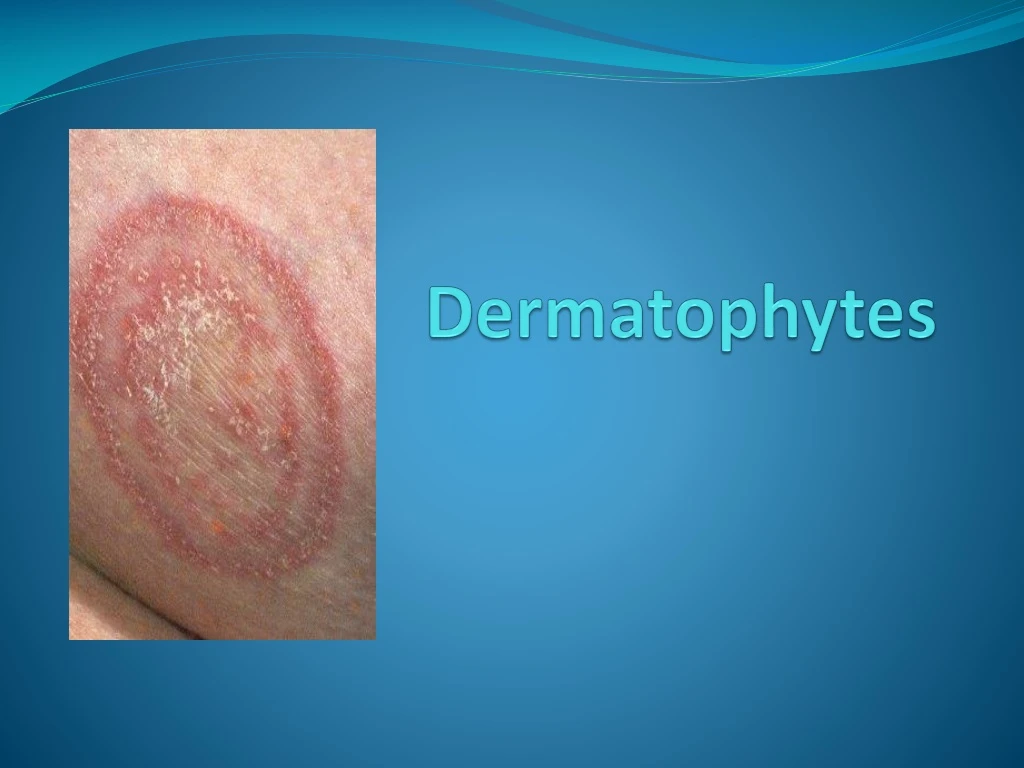
Pathogenesis: • Dermatophytes grow only within dead, keratinized tissue • The fungal cells produce keratinolytic proteases • Fungal metabolic products diffuse into the epidermis to cause erythema, vesicles, pustules along with pruritis • Hyphae become old, break into arthrospores which are shed off in time • This is partially responsible for the central clearing in ringworm type of lesions
The hyphal tips growing down within hair shaft reach the edges of living keratinizing cells and form Adamson’s fringe • The infective process ceases and healing occurs when the balance of fungus and host is tilted in favour of host • Clinical features result from combination of keratin destruction and inflammatory response generated by host
Clinical features • Ringworm or tinea depending on the anatomical site involved • Term ‘tinea’ is derived from latin word meaning ‘worm’ or ‘moth’ • inflammation is seen maximum at the advancing margins leaving central area with some clearing
Tineacapitis: • Infection of shaft of scalp hairs • Kerion: severely painful inflammatory reaction producing raised, circumscribed boggy mass on scalp, with suppurative lesions on the scalp, discharging pus • Favus: condition with cup-like crusts around infected follicles, may lead to patchy alopecia and scarring
Ectothrix infection: hyphae produce arthrospores arranged as mosaic sheath around hair or as chains on surface of hair shaft, • cuticle of hair remains intact
Endothrix infection: hyphae form arthrospores within hair shaft, which is severely weakened • cuticle of hair is usually destroyed
Tineacorporis: disease of the skin over body, result from extension of infection from scalp, groin or beard • Erythematous scaly lesions, annular, sharply marginated plaques with raised border which may be single or multiple
Tineagladiotorum: is an emerging infection in wrestlers • It is as a result of direct skin- to-skin contact • Lesions are on arms, trunk or head and neck • Tineafaciei: infection of non-bearded regions of face • Tineacruris: infection of groin and mostly present in men using tight-fitting garments • Seen in perineum, scrotum, perianal area
Tineabarbae: infection of beard and moustache areas of face, erythematous patches on face which show scaling, fragile lusterless hairs
Tineamanuum: infection of skin of palmar aspect of hands • Diffuse hyperkeratosis of palms and fingers • Tineapedis: infection of plantar aspect of foot, toes and interdigital web spaces • Warmth and moisture produced by shoes are key factors in infection • Seen in people wearing shoes for long hours • Popularly known as Athlete’s foot • Scaling, fissuring, erythema with itching or burning sensation • Small vesicles rupture and discharge thin fluid
Tineaunguium: infection of nail plates • Begins with the free edge of nail plate or along lateral nail fold and may continue until entire nail plate or nail bed is infected • Onychomycosis: fungal infection of nail- dermatophytes or non-dermatophytes
Id reaction: Dermatophytid reaction: • Secondary eruption occuring in sensitisedtinea patients because of ciculation of allergenic products from primary site of infection • This may develop following the initiation of oral antifungal therapy • Frequently seen in patients with absence of delayed reaction to dermatophytic antigen i.e. trichophytin
Laboratory diagnosis • Specimen collected: • Skin scrapings, nail clippings, plucked infected hair, pus etc • Selection of infected hair for examination by Wood’s lamp • Microscopy: • 10% KOH wet mount examination: fungus appears as branching hyaline mycelia, with arthrospores • Examination of hair for endothrix, ectothrix • Examination of nails, skin
Fungal culture: • On Sabouraud’s dextrose agar • Species identification by culture examination- macroscopic appearance and microscopic appearance • Lactophenol cotton blue stain (LP mount) for microscopic examination • Growth is slow and colonies appear only in 1-3 weeks
Trichophyton: Colonies are powdery, velvety or waxy with pigmentation characteristic of species • Microconidia are abundant and arranged in clusters along the hyphae or borne on conidiophores • Macroconidia are relatively scanty
Microsporum: • Colonies are cottony, velvety white to brown pigmentation • Microconidia are relatively scanty • Macroconidiaare predominant, large, multicellular, spindle-shaped
Epidermophyton: • Colonies are powdery and greenish yellow (khaki-coloured), with raised and folded centre • Microconidia are absent • Macroconidiaare multi-cellular, pear-shaped and arranged in clusters
Skin infection Hair infection Nail infection • Trichophyton • Microsporum • Epidermophyton Hair infection Skin infection Skin infection Nail infection
Treatment: • Topical antifungal agents are effective for skin infections • Oral griseofulvin is the drug of choice