Download
1 / 60
670 likes | 932 Views
Liver Cirrhosis. K. Dionne Posey, MD, MPH Internal Medicine & Pediatrics December 9, 2004. Introduction. The two most common causes in the United States are alcoholic liver disease and hepatitis C, which together account for almost one-half of those undergoing transplantation. Introduction.

E N D
Liver Cirrhosis K. Dionne Posey, MD, MPH Internal Medicine & Pediatrics December 9, 2004
Introduction • The two most common causes in the United States are alcoholic liver disease and hepatitis C, which together account for almost one-half of those undergoing transplantation
Introduction • 12th leading cause of death in the united states in2002 • On average about 27,000 deaths per year • Patients with cirrhosis are susceptible to a variety of complications and their life expectancy is markedly reduced
Exactly How Much Do You Drink? • Estimated that the development of cirrhosis requires, on average, the ingestion of 80 grams of ethanol daily for 10 to 20 years • This corresponds to approximately one liter of wine, eight standard sized beers, or one half pint of hard liquor each day
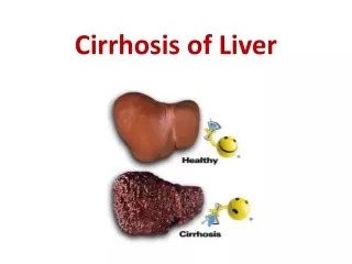
Pathophysiology • Irreversible chronic injury of the hepatic parenchyma • Extensive fibrosis - distortion of the hepatic architecture • Formation of regenerative nodules
Clinical Manifestations • Spider angiomas • Palmar erythema • Nail changes • Muehrcke's nails • Terry’s nails • Gynecomastia • Testicular atrophy
Muehrcke's nails Terry’s nails Clinical Manifestations
Fetor hepaticus Jaundice Asterixis Pigment gallstones Parotid gland enlargement Cruveilhier-Baumgarten murmur Hepatomegaly Splenomegaly Caput medusa Clinical Manifestations
Laboratory Studies • most common measured laboratory test classified as LFTs include • the enzyme tests (principally the serum aminotransferases, alkaline phosphatase, and gamma glutamyl transpeptidase), the serum bilirubin • tests of synthetic function (principally the serum albumin concentration and prothrombin time)
Radiologic Modalities • Can occasionally suggest the presence of cirrhosis, they are not adequately sensitive or specific for use as a primary diagnostic modality • Major utility of radiography in the evaluation of the cirrhotic patient is in its ability to detect complications of cirrhosis
Diagnosis • Liver biopsy • Obtained by either a percutaneous, transjugular, laparoscopic, or radiographically-guided fine-needle approach • Sensitivity of a liver biopsy for cirrhosis is in the range of 80 to 100 percent depending upon the method used, and the size and number of specimens obtained
Diagnosis • not necessary if the clinical, laboratory, and radiologic data strongly suggest the presence of cirrhosis • liver biopsy can reveal the underlying cause of cirrhosis
Morphologic Classification • Micronodular cirrhosis • Nodules less than 3 mm in diameter • Believed to be caused by alcohol, hemochromatosis, cholestatic causes of cirrhosis, and hepatic venous outflow obstruction
Morphologic Classification • Macronodular cirrhosis • Nodules larger than 3 mm • Believed to be secondary to chronic viral hepatitis
Morphologic Classification • Relatively nonspecific with regard to etiology • The morphologic appearance of the liver may change as the liver disease progresses • micronodular cirrhosis usually progresses to macronodular cirrhosis • Serological markers available today are more specific than morphological appearance of the liver for determining the etiology of cirrhosis • Accurate assessment of liver morphology may only be achieved at surgery, laparoscopy, or autopsy
Complications • Ascites • Spontaneous Bacterial Peritonitis • Hepatorenal syndrome • Variceal hemorrhage • Hepatopulmonary syndrome
Complications • Other Pulmonary syndromes • Hepatic hydrothorax • Portopulmonary HTN • Hepatic Encephalopathy • Hepatocellular carcinoma
Ascites • Accumulation of fluid within the peritoneal cavity • Most common complication of cirrhosis • Two-year survival of patients with ascites is approximately 50 percent
Ascites • Assessment of ascites • Grading • Grade 1 — mild; Detectable only by US • Grade 2 — moderate; Moderate symmetrical distension of the abdomen • Grade 3 — large or gross asites with marked abdominal distension • Older system -subjective • 1+ minimal, barely detectable • 2+ moderate • 3+ massive, not tense • 4+massive and tense
Ascites • Imaging studies for confirmation of ascites • Ultrasound is probably the most cost-effective modality
Ascites • Treatment aimed at the underlying cause of the hepatic disease and at the ascitic fluid itself • Dietary sodium restriction • Limiting sodium intake to 88 meq (2000 mg) per day
Ascites • The most successful therapeutic regimen is the combination of single morning oral doses of Spironolactone and Furosemide, beginning with 100 mg and 40 mg • Two major concerns with diuretic therapy for cirrhotic ascites: • Overly rapid removal of fluid • Progressive electrolyte imbalance
Spontaneous Bacterial Peritonitis • Infection of ascitic fluid • Almost always seen in the setting of end-stage liver disease • The diagnosis is established by • A positive ascitic fluid bacterial culture • Elevated ascitic fluid absolute polymorphonuclear leukocyte (PMN) count ( >250 cells/mm3)
Spontaneous Bacterial Peritonitis • Clinical manifestations: • Fever • Abdominal pain • Abdominal tenderness • Altered mental status
Hepatorenal syndrome • acute renal failure coupled with advanced hepatic disease (due to cirrhosis or less often metastatic tumor or severe alcoholic hepatitis) • characterized by: • Oliguria • benign urine sediment • very low rate of sodium excretion • progressive rise in the plasma creatinine concentration
Hepatorenal Syndrome • Reduction in GFR often clinically masked • Prognosis is poor unless hepatic function improves • Nephrotoxic agents and overdiuresis can precipitate HRS
Variceal hemorrhage • Occurs in 25 to 40 percent of patients with cirrhosis • Prophylactic measures • Screening EGD recommended for all cirrhotic patients
Hepatopulmonary syndrome • Hepatopulmonary syndrome • Liver disease • Increased alveolar-arterial gradient while breathing room air • Evidence for intrapulmonary vascular abnormalities, referred to as intrapulmonary vascular dilatations (IPVDs)
Hepatic Hydrothorax • Pleural effusion in a patient with cirrhosis and no evidence of underlying cardiopulmonary disease • Movement of ascitic fluid into the pleural space through defects in the diaphragm, and is usually right-sided • Diagnosis -pleural fluid analysis • reveals a transudative fluid • serum to fluid albumin gradient greater than 1.1
Hepatic hydrothorax • Confirmatory study: • Scintigraphic studies demonstrate tracer in the chest cavity after injection into the peritoneal cavity • Treatment options: • diuretic therapy • periodic thoracentesis • TIPS
Portopulmonary HTN • Refers to the presence of pulmonary hypertension in the coexistent portal hypertension • Prevalence in cirrhotic patients is approximately 2 percent • Diagnosis: • Suggested by echocardiography • Confirmed by right heart catheterization
Hepatic Encephalopathy • Spectrum of potentially reversible neuropsychiatric abnormalities seen in patients with liver dysfunction • Diurnal sleep pattern pertubation • Asterixis • Hyperactive deep tendon reflexes • Transient decerebrate posturing
Hepatic Encephalopathy • Monitoring for events likely to precipitate HE [i.E.- variceal bleeding, infection (such as SBP), the administration of sedatives, hypokalemia, and hyponatremia] • Reduction of ammoniagenic substrates • Lactulose / lactitol • Dietary restriction of protein • Zinc and melatonin
Hepatocellular Carcinoma • Patients with cirrhosis have a markedly increased risk of developing hepatocellular carcinoma • Incidence in well compensated cirrhosis is approximately 3 percent per year
Hepatocellular Carcinoma • Symptoms are largely due to mass effect from the tumor • Pain, early satiety, obstructive jaundice, and a palpable mass • Serum AFP greater than 500 micrograms/l in a patient with cirrhosis are virtually diagnostic • Median survival following diagnosis is approximately 6 to 20 months
Prognostic Tools • MELD (model for end-stage liver disease) • Identify patients whose predicted survival post-procedure would be three months or less • MELD = 3.8[serum bilirubin (mg/dL)] + 11.2[INR] + 9.6[serum creatinine (mg/dL)] + 6.4
Prognostic Tools • Child-Turcotte-Pugh (CTP) score • initially designed to stratify the risk of portacaval shunt surgery in cirrhotic patients • based upon five parameters: serum bilirubin, serum albumin, prothrombin time, ascites and encephalopathy • good predictor of outcome in patients with complications of portal hypertension