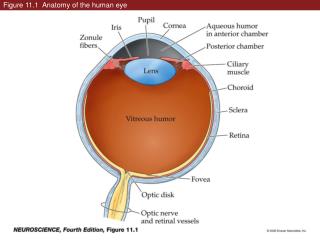
Download
1 / 43
430 likes | 534 Views
Figure 30.1 Sexually dimorphic anatomy in the hawk moth, Manduca sexta. Figure 30.1 Sexually dimorphic anatomy in the hawk moth, Manduca sexta (Part 1). Figure 30.1 Sexually dimorphic anatomy in the hawk moth, Manduca sexta (Part 2).

E N D
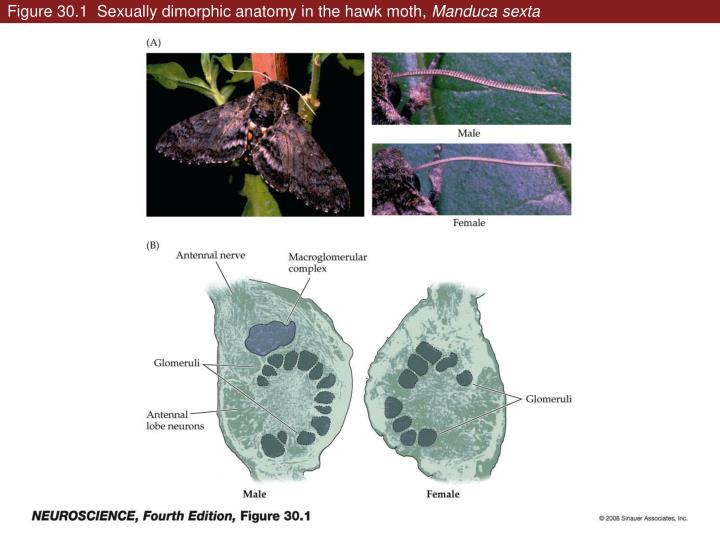
Figure 30.1 Sexually dimorphic anatomy in the hawk moth, Manduca sexta
Figure 30.1 Sexually dimorphic anatomy in the hawk moth, Manduca sexta (Part 1)
Figure 30.1 Sexually dimorphic anatomy in the hawk moth, Manduca sexta (Part 2)
Figure 30.2 Chromosomal sex and primary sex determination in humans
Figure 30.3 Gonadal sex steroids and their organizational influence
Figure 30.3 Gonadal sex steroids and their organizational influence (Part 1)
Figure 30.3 Gonadal sex steroids and their organizational influence (Part 2)
Figure 30.5 Sex differences in innervation of the perineal muscles
Figure 30.5 Sex differences in innervation of the perineal muscles (Part 1)
Figure 30.5 Sex differences in innervation of the perineal muscles (Part 2)
Figure 30.5 Sex differences in innervation of the perineal muscles (Part 3)
Figure 30.6 Hypothalamic nuclei are sexually dimorphic and their neuronal activity is associated with sexual behaviors
Figure 30.6 Hypothalamic nuclei are sexually dimorphic and their neuronal activity is associated with sexual behaviors (Part 1)
Figure 30.6 Hypothalamic nuclei are sexually dimorphic and their neuronal activity is associated with sexual behaviors (Part 2)
Figure 30.7 Hypothalamic regulation of lactation in nursing mothers
Figure 30.7 Hypothalamic regulation of lactation in nursing mothers (Part 1)
Figure 30.7 Hypothalamic regulation of lactation in nursing mothers (Part 2)
Figure 30.8 Cortical representation of the chest wall in the rat primary somatic sensory cortex during lactation
Figure 30.8 Cortical representation of the chest wall in the rat primary somatic sensory cortex during lactation (Part 1)
Figure 30.8 Cortical representation of the chest wall in the rat primary somatic sensory cortex during lactation (Part 2)
Figure 30.8 Cortical representation of the chest wall in the rat primary somatic sensory cortex during lactation (Part 3)
Figure 30.8 Cortical representation of the chest wall in the rat primary somatic sensory cortex during lactation (Part 4)
Figure 30.9 Estrogen and testosterone influence neuronal growth and differentiation
Figure 30.9 Estrogen and testosterone influence neuronal growth and differentiation (Part 1)
Figure 30.9 Estrogen and testosterone influence neuronal growth and differentiation (Part 2)
Figure 30.9 Estrogen and testosterone influence neuronal growth and differentiation (Part 3)
Figure 30.10 Estrogen influences synaptic transmission (Part 1)
Figure 30.10 Estrogen influences synaptic transmission (Part 2)
Figure 30.11 Distribution in the rat brain of the three major receptor/transcription factors that bind sex hormones
Figure 30.11 Distribution in the rat brain of the three major receptor/transcription factors that bind sex hormones (Part 1)
Figure 30.11 Distribution in the rat brain of the three major receptor/transcription factors that bind sex hormones (Part 2)
Figure 30.12 Sex-specific splice isoforms of the Drosophila fruitless gene correlate with sex-specific courtship and mating behaviors
Figure 30.12 Sex-specific splice isoforms of the Drosophilafruitless gene correlate with sex-specific courtship and mating behaviors (Part 1)
Figure 30.12 Sex-specific splice isoforms of the Drosophila fruitless gene correlate with sex-specific courtship and mating behaviors (Part 2)
Figure 30.12 Sex-specific splice isoforms of the Drosophila fruitless gene correlate with sex-specific courtship and mating behaviors (Part 3)
Figure 30.13 Distinct patterns of activation of estrogen and androgen in women and men
Figure 30.14 Brain regions that differ in size in females versus males
Figure 30.15 Sex-specific activation of the amygdala in response to memory of images with defined emotional content