Download
1 / 4
40 likes | 123 Views
This study delves into the analysis of the ATP9.5' trailer and promoter region through PCR techniques. By closely examining the ATP9 pre-mRNA and mRNA sequences, along with the 5' leader regions, the study sheds light on the cleavage and endonucleolytic sites of the ATP9 gene. The use of modified CR-RT-PCR with 5S rRNA as an adapter molecule helps determine the 3' end of the ATP9.5' trailer. The results provide valuable insights into the molecular mechanisms governing gene expression in this region.

E N D
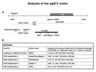
Analysis of the atp9 5‘ trailer A promoter atp9 3’ 5’ (-239) atp9 pre-mRNA (+180) atp9 mRNA (-84/-83) atp9 5’ leader Atatp9.Endo.Mega.A Atatp9-11 5S rRNA atp9 5’ leader At5S-1 At5S-Mega.R At5S-5 B
C D 2. PCR; on template II 2. PCR; on template I marker 1. PCR marker 1 marker 2 2,000 bp 2,000 bp 1,500 bp 1,500 bp 1,000 bp 1,000 bp I 750 bp 750 bp 500 bp 500//501/489 bp 404 bp 331 bp 250 bp II 250//242 bp III 190 bp 147 bp 111/110 bp E
F */** A single* or two** nucleotide(s) at the ligation site could be either assigned to atp9 or 5S rRNA. Thus the atp9 5’-leader ends either at the indicated position or one or two nucleotide(s) further downstream.
Supplementary Figure 28. Analysis of the atp9 5‘ trailer. A modified CR-RT-PCR using endogenous 5S rRNA as adapter molecule was carried out to determine the 3’ end of the atp9 5’ trailer upstream of the mature atp9 transcript as outlined in (A). The site of the endonucleolytic cleavage is marked by a pair of scissors. The primers used are listed in table (B). The products of the first PCR amplification (marked by arrows and detailed in table (E)) were separatedbyagarose gel electrophoresis (C), excised and used as template for a second PCR amplification. PCR product I was additionally analyzed by direct sequencing. The products of the second PCR were again separated by agarose gel electrophoresis (D). PCR product III was cloned and individual clones were sequenced. The ends found in these clones are listed in table (F).