Download
1 / 95
970 likes | 1.24k Views
Tissue: The Living Fabric. P A R T A. Tissues. Groups of cells similar in structure and function The four types of tissues Epithelial Connective Muscle Nervous. Epithelial Tissue. Cellularity – composed almost entirely of cells

E N D
Tissue: The Living Fabric P A R T A
Tissues • Groups of cells similar in structure and function • The four types of tissues • Epithelial • Connective • Muscle • Nervous
Epithelial Tissue • Cellularity – composed almost entirely of cells • Form continuous sheets held together by tight junctions and desmosomes • Polarity – apical and basal surfaces
Epithelial Tissue • Attached to a basement membrane • Lamina lucida (basal) • Lamina densa (reticular) • Avascularbut innervated – contains no blood vessels but supplied by nerve fibers • Regenerative – rapidly replaces lost cells by cell division
Classification of Epithelia Figure 4.1a
Classification of Epithelia Figure 4.1b
Epithelia: Simple Squamous • Single layer of flattened cells • Disc-shaped nuclei • Sparse cytoplasm • Functions • Diffusion and filtration • Provide a slick, friction-reducing lining in lymphatic and cardiovascular systems
Epithelia: Simple Squamous • Present in the kidney glomeruli, lining of heart, blood vessels, lymphatic vessels, and serosae
Epithelia: Simple Cuboidal • Single layer of cube-like cells • Spherical central nuclei • Function in secretion and absorption • Present in kidney tubules, ducts and secretory portions of small glands, and ovary surface
Epithelia: Simple Columnar • Single layer of tall cells • Oval nuclei; many contain cilia • Goblet cells are often found in this layer • Function in absorption and secretion • Nonciliated type line digestive tract and gallbladder
Epithelia: Simple Columnar • Ciliated type line small bronchi, uterine tubes, and some regions of the uterus • Cilia help move substances through internal passageways
Epithelia: Pseudostratified Columnar • Single layer of cells with different heights; some do not reach the free surface • Nuclei are seen at different layers • Function in secretion and propulsion of mucus • Present in the male sperm-carrying ducts (nonciliated) and trachea (ciliated)
Epithelia: Stratified Squamous • Thick membrane composed of several layers of cells • Function in protection of underlying areas subjected to abrasion • Forms the external part of the skin’s epidermis (keratinized cells), and linings of the esophagus, mouth, and vagina (nonkeratinized cells)
Epithelia: Stratified Cuboidal and Columnar • Stratified cuboidal • Quite rare in the body • Found in some sweat and mammary glands • Typically two cell layers thick
Epithelia: Stratified Cuboidal and Columnar • Stratified columnar • Limited distribution in the body • Found in the male urethra, and lining some glandular ducts • Also occurs at transition areas between two other types of epithelia
Epithelia: Transitional • Several cell layers, basal cells are cuboidal, surface cells are dome shaped • Stretches to permit the distension of the urinary bladder • Lines the urinary bladder, ureters, and part of the urethra
Epithelia: Glandular • A gland is one or more cells that produces secretion • Classified by: • Site of secretion release • Endocrine • Exocrine • Number of cells forming the gland • Unicellular • Goblet cell • Multicellular
Endocrine Glands • Ductless glands • The product is secreted into the interstitial fluid • Hormones • Secretions include amino acids, proteins, glycoproteins, and steroids
Exocrine Glands • More numerous than endocrine glands • Secrete their products onto body surfaces (skin) or into body cavities • Examples include mucous, sweat, oil, and salivary glands • Multicellular exocrine glands are composed of a duct and secretory unit
GobletCell Figure 4.3b
Structural Classification of Multicellular Exocrine Glands Figure 4.4a–d
Structural Classification of Multicellular Exocrine Glands Figure 4.4e–g
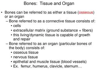
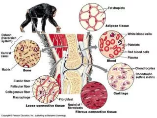
Connective Tissue • Found throughout the body; most abundant and widely distributed in primary tissues • Embryonic • Mesenchyme • Adult
Functions of Connective Tissue • Binding and support • Protection • Insulation • Transportation
Characteristics of Connective Tissue • Connective tissues have: • Mesenchyme as their common tissue of origin • Varying degrees of vascularity • Nonliving extracellular matrix • ground substance • fibers • cells
Structural Elements of Connective Tissue • Ground substance – unstructured material that fills the space between cells • Fibers – collagen, elastic, or reticular • Cell population – fibroblasts, chondroblasts, osteoblasts, etc
Ground Substance Composition • Interstitial (tissue) fluid • Adhesion proteins – fibronectin and laminin. Attaches cells to matrix components • Proteoglycans – glycosaminoglycans (GAGs) • Functions as a molecular sieve through which nutrients diffuse between blood capillaries and cells
Ground Substance: Proteoglycan Structure Figure 4.7
Fibers • Collagen – tough; provides high tensile strength. White fibers • Elastic – long, thin fibers that allow for stretch. Yellow fibers • Reticular – branched collagenous fibers that form delicate networks
Cells • Fibroblasts – connective tissue proper • Chondroblasts – cartilage • Osteoblasts – bone • Hematopoietic stem cells – blood • White blood cells, plasma cells, macrophages, and mast cells • Adipocytes
Connective Tissue: Embryonic • Mesenchyme – embryonic connective tissue • Gel-like ground substance with fibers and star-shaped mesenchymal cells • Gives rise to all other connective tissues • Found in the embryo
Connective Tissue Proper: Loose • Areolar connective tissue • Gel-like matrix with all three connective tissue fibers • Fibroblasts, macrophages, mast cells, and some white blood cells • Wraps and cushions organs • Widely distributed throughout the body • Well vascularized
Areolar Connective Tissue Figure 4.8
Connective Tissue Proper: Loose • Adipose connective tissue • Closely packed adipocytes • Well vascularized • Found under skin, around kidneys, within abdomen, etc • Local fat deposits serve nutrient needs of highly active organs • White fat vs. brown fat
Connective Tissue Proper: Loose • Reticular connective tissue • Loose ground substance with reticular fibers • Reticular cells lie in a fiber network • Forms a soft internal skeleton, or stroma, that supports other cell types • Found in lymph nodes, bone marrow, and the spleen