Download
1 / 17
170 likes | 189 Views
Explore innovative eye plaques for conformal treatment planning of intraocular tumors using cutting-edge technologies. Learn how the Plaque Simulator™ aids in precise positioning and customizable solutions.

E N D
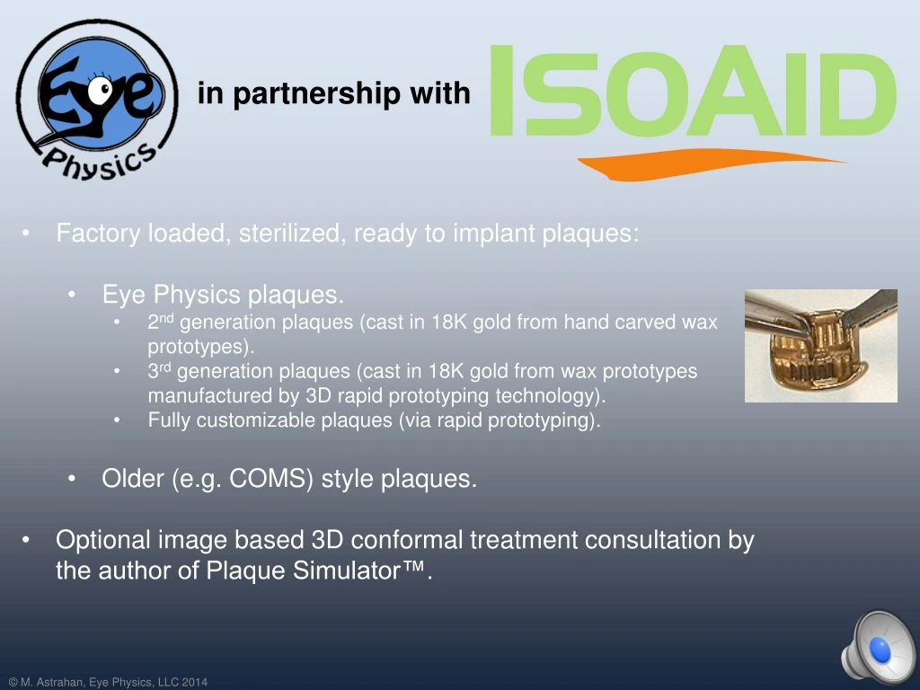
Factory loaded, sterilized, ready to implant plaques: • Eye Physics plaques. • 2nd generation plaques (cast in 18K gold from hand carved wax prototypes). • 3rd generation plaques (cast in 18K gold from wax prototypes manufactured by 3D rapid prototyping technology). • Fully customizable plaques (via rapid prototyping). • Older (e.g. COMS) style plaques. • Optional image based 3D conformal treatment consultation by the author of Plaque Simulator™. in partnership with
CT and MR imaging alone do not have the resolution required for conformal treatment of intraocular tumors. • A method for delivering conformal treatment is also required. Image Based Conformal Treatment Planning for Intraocular Tumors
Conformal planning of intraocular tumors requires a 3D fusion of imaging modalities including fundus photograhy, CT (or MR), and ultrasound. Image Based Conformal Treatment Planning for Intraocular Tumors Fundus Photos CT Ultrasound 3D Model + + =
Image based planning for 3D conformal treatment of intraocular tumors using eye plaques is not new. • The software component (Plaque Simulator™) was developed by Eye Physics founder Prof. Emeritus Melvin Astrahan, PhD, DABR at the University of Southern California (USC) Keck School of Medicine ca. 1990. • Plaque Simulator™ was licensed from USC for international commercial distribution in 1994. • There are many users of Plaque Simulator™ throughout the world. Image Based Conformal Treatment Planning for Intraocular Tumors
A collage of fundus photographs is used to map the tumor base and determine the location of the base with respect to easily identifiable retinal landmarks. How Fundus Photography is Used Fundus camera photo courtesy of NIDEK Inc. • The fundus collage can be calibrated by finding these same landmarks in 3D CT space. • However, fundus photos do not provide the tumor height. • Fundus photos are also not possible for anterior (e.g. ciliary body) tumors. Tumor Optic Disc Posterior Pole (above the fovea)
The 3D coordinates of the posterior pole on the retina and the center of the optic disc can be closely approximated from an axial CT (or MR) image which bisects the eye through the optic nerve. How CT and MR Images are Used • CT (or MR) images also provide the location of the limbus, the equatorial diameter, and the oblate curvature of the eye. • Fundus photos can be calibrated by finding the posterior pole and optic disc in the fundus collage. • A 3D model of the fused images can be created. limbus posterior pole optic disc optic nerve
2D ultrasound (US) images provide the best measurement of tumor height. • US images help differentiate between tumor and retinal detachment. • US is essential for anterior tumors (e.g. ciliary) that cannot be photographed. How Ultrasound Images are Used • US images, however, do not show the shape of the tumor base and are difficult to fuse with CT and fundus photos because it is difficult to find common points of reference.
Historically, plaques have assumed simple shapes (e.g. circular perimeters) because the precise shape of the tumor base and its location within the eye were not available for advance treatment planning. These details were often not precisely determined until the time of surgery. • At surgery, the tumor base was marked by transilluminating the eye, a shadow of the tumor was cast onto the sclera, the shadow was outlined with ink, and then we hoped that the prepared circular plaque was large enough to cover the tumor base and dosimetric margin. How Plaques Were Surgically Positioned • Nonconformal preplanning can lead to either the prescribed tumorcidal dose being delivered to a larger portion of the retina surrounding the tumor base than may be necessary to assure tumor control, or portions of the tumor receiving a less than prescribed dose if the plaque was too small. • Shadow casting can lead to plaque positioning errors depending on the position of the light source and the tumor height and location. • Suturing a small diameter plaque over a small tumor at the posterior of the eye is very difficult.
The tumor is digitized by projecting the calibrated fundus collage onto the retinal diagram, a polar map of the retina. • A plaque that fits the tumor and ocular anatomy is selected. • Surgical coordinates for the plaque eyelets are calculated and expressed as clock hour and caliper distance from the limbus on the retinal diagram. How Intraocular Tumors are Located in Plaque Simulator
The surgeon marks the meridian planes on the sclera at the limbus using a toric axis marker and then uses a Castroviejo caliper to mark the suture point on the meridian. • Placement uncertainty is about 0.5 mm so PTV margins of 2 mm surrounding the tumor base are usually sufficient. How the Plaque is Positioned Using The PS Coordinate System • The distance between the suture eyelets is used to cross-check the meridian spacing.
Seed positions in the plaque are then selected to conform to the tumor base and spare the macula and optic disc as much as possible. How Conformal Dosimetry is Reliably Achieved • Conformal treatment can only be reliably delivered if the surgeon uses the PS suture eyelet coordinates (which, fortunately, they are already familiar with). • For extremely posterior tumors, the eyelets of an elliptical and/or notched plaque can be positioned on the anterior hemisphere of the eye where they are easier to suture.
Plaque Simulator displays radiation dose in various 2D formats such as meridian and equatorial planes, and on the retinal surface to assure tumor coverage and reduce dose to critical regions such as the macula. • 3D displays are also available. • Collimation of primary radiation by the gold shell and attenuation in the Silastic™ carrier of COMS style plaques are accounted for. • Accounting for primary radiation is a good approximation because most attenuation at I-125 energies is photoelectric in nature. • Plaque Simulator uses the AAPM TG43 methodology with plaque specific enhancements derived from Monte Carlo modeling and physical measurement. How Conformal Dosimetry is Calculated
The implant is simulated using 3D interactive graphics. The surgeon will know, before beginning the implant, the coordinates of every suture and which muscles may interfere with the plaque. How Conformal Treatment is Simulated • Tumor and PTV coverage are assured because the tumor dimensions are accurately determined in the planning process. • This saves time in the OR and prevents recurrences which might result from incorrect plaque size or uncertainty in plaque positioning.
Eye Physics plaques simplify both dosimetry (no Silastic attenuation) and surgery compared to other designs. • Eye Physics plaques are thinner (<= 2mm), have larger, more rugged suture eyelets, can have deep notches to treat tumors near the optic disc, and can be flash sterilized. • The plaques and software have a 25 year history of successful clinical use at USC. • Each radiation source in an Eye Physics plaque is mounted in a collimating slot. Retina adjacent to the plaque is protected from most laterally directed primary radiation. • There is no point to vitreous fluid replacement with higher (than water) atomic number oils when Eye Physics plaques are used. COMS Eye Physics The Eye Physics Plaques
Every seed in a COMS plaque irradiates the retina and sclera under the tumor and adjacent to the plaque. • Retinal dose at the base of the tumor can be up to 10X the dose prescribed to the apex of medium to large tumors. • COMS plaque dosimetry is complicated by the atomic composition of the silicone seed carrier for low energy x-rays. • COMS plaques are thick at the edges and are designed to fit a spherical eye of one radius of curvature. • Eye Physics plaques collimate the radiation from each seed to remove lateral radiation that does not contribute to the tumor dose. • Retinal dose at the base of the tumor is usually only 2X to 3X the dose prescribed to the apex of medium to large tumors. • Eye Physics plaques are designed using Plaque Simulator technology and are manufactured using computer aided rapid prototyping technology. • Eye Physics plaques are <= 2mm thick and are available in many shapes, sizes and even mixed curvatures to perfectly fit real eyes.
These wax prototypes were designed using Plaque Simulator software and manufactured using 3D rapid prototyping technology to precisely match the Plaque Simulator models. The wax prototypes are then cast in gold to make the final product. Eye Physics 3rd generation computer designed and prototyped plaques will soon be available in dozens of preconfigured as well as fully customizable sizes, shapes, curvatures and source arrangements. Our plaques are <= 2 mm thick and the radionuclide sources are individually collimated to create a dose distribution which conforms to the tumor, spares the surrounding retina, and provides a near perfect mechanical fit to each patient's eye.
Luxton G, Astrahan MA and Petrovich Z. Measurements of backscatter from a single seed of I-125 for eye plaque dosimetry. Medical Physics 15:397-400, 1988. Luxton G, Astrahan MA, Liggett P, Neblett DL, Cohen DM and Petrovich Z. Dosimetric calculations and measurements of gold plaque ophthalmic irradiators using Iridium-192 and Iodine-125 seeds. International Journal of Radiation Oncology, Biology, Physics 15:167-176, 1988. Astrahan M, Luxton G, Jozsef G, Kampp TD, Liggett P and Sapozink MD. An interactive treatment planning system for ophthalmic plaque radiotherapy. International Journal of Radiation Oncology, Biology, Physics 18:679-687, 1990. Luxton G, Astrahan M, Findley D and Petrovich Z. Measurement of dose rate from exposure-calibrated I-125 seeds. International Journal of Radiation Oncology, Biology, Physics 18:1199-1207, 1990. Lean EK, Cohen D, Liggett P, Luxton G, Hyden E, Green R, Astrahan MA and Petrovich Z. Episcleral radioactive plaque therapy: initial clinical experience with 56 patients. Amer. J. Clin. Oncology 13:185-190,1990. Petrovich Z, Liggett PE, Luxton G, Lean E, Langholz B and Astrahan MA. Radioactive plaque therapy in the management of primary malignant ocular melanoma: An overview. Endocurietherapy, Hyperthermia Oncology, 6: 131-141, 1990. Astrahan M, Luxton G, Jozsef G, Liggett P and Petrovich Z. Optimization of I-125 ophthalmic plaque brachytherapy. Medical Physics 17:1053-1057, 1990. Liggett PE, Ma C, Astrahan MA, Pince KJ, Green R, McDonnell J and Petrovich Z. Combined localized current field hyperthermia and irradiation for intraocular tumors. Ophthalmol 98:1830-1836, 1991. Petrovich Z, Astrahan M, Luxton G, Green R, Langholz B and Liggett P. Episcleral plaque thermoradiotherapy in patients with choroidal melanoma. International Journal of Radiation Oncology, Biology, Physics 23:599-603, 1992. Petrovich Z, Luxton G, Langholz B, Astrahan MA and Liggett PE. Episcleral plaque radiotherapy in the treatment of uveal melanomas. Int J Radiat Oncol Biol Phys 24:247-251, 1992. Evans MDC, Astrahan MA, and Bate R. Tumor localization using fundus view photography for episcleral plaque therapy. Medical Physics 20:769-775, 1993. Petrovich Z, Pike M, Astrahan MA, Luxton G, Murphree AL and Liggett PE. Episcleral plaque thermoradiotherapy. Am J Clin Oncol 19:207-211, 1996. Astrahan MA, Luxton G, Pu Q and Petrovich Z. Conformal Episcleral Plaque Therapy. International Journal of Radiation Oncology, Biology, Physics 39:505-519, 1997. Astrahan M. A patch source model for treatment planning of ruthenium ophthalmic applicators. Medical Physics 30(6):1219-1228, 2003. Astrahan M. Improved treatment planning for COMS eye plaques, International Journal of Radiation Oncology, Biology, Physics,61(4):1227-1242, 2005. Astrahan M, Szechter A and Finger P. Design and Dosimetric Considerations of a Modified COMS Plaque: The reusable seed-guide insert. Medical Physics 32(8), 2706-2716, 2005.