Download

1 / 14
140 likes | 225 Views
Incontinentia Pigmenti (IP) is an X-linked genodermatosis primarily affecting females, characterized by distinct dermatological stages and associated with NEMO gene mutations. It presents skin abnormalities and may also involve eye and central nervous system anomalies. This condition is caused by NEMO genomic rearrangement, influencing NFkB signaling and X chromosome inactivation. Understanding the XCI process is crucial for grasping the mechanisms underlying IP development.

E N D
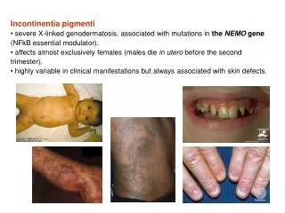
Incontinentiapigmenti (IP) • X-linked genodermatosis, associated with mutations in the NEMOgene(NFkB essential modulator, Xq28) • Affects almost exclusively females (males die in utero before the second trimester) • Highly variable in clinical manifestations but always associated with skin defects
Incontinentiapigmenti (IP) - characterized by four distinct dermatological stages that begin within 2 weeks after birth with blisters and inflammatory response (Stage I/Vesicular Stage, Fig. 1). Subsequently, verrucous hyperkeratotic lesions develop (Stage II/Verrucous Stage, Fig. 2) and disappear over time, leaving areas of hyperpigmentation due to melanin accumulation (Stage III/Hyperpigmented Stage, Fig. 3). These areas generally disappear by the second decade, but adults may still show areas of dermal scarring. Fig. 1 Fig. 2 Fig. 3
Incontinentiapigmenti (IP) • Every IP patient exhibits skin abnormalities,blindness and central nervous system anomalies are occurring in about 40% and 30% of patients, resp. • Eye manifestations in IP patients (retinal detachment and consequent blindness) ← deficient vascularization of retina. • CNS manifestations in IP patients (ischemia, atrophy, seizures, paralysis, mental retardation) ← deficient vascularization of brain. • In addition to the dermal, visual and brain defects, IP patients exhibit some less medically significant problems, including hair loss (alopecia); conical, peg-shaped or absent teeth (anodontia), and nail dystrophy.
Incontinentiapigmenti (IP), NEMO GenomicrearrangementofNEMO • Xq28, the NEMO gene. • IP rearrangement - excisionofthe region betweentwo MER67B repeatslocatedupstreamof exon 4 and downstreamof exon 10. Multiplex PCR products in IP patients and controls. Presence of 1045 bp band indicates the presence of the common rearrangement in IP patients. 733 bp product serves as internal amplification control. G Courtois, Cell Death and Differentiation (2006)
NEMO, NFkB • Resting cells: NFkB is kept inactive in cytoplasm through interaction with IkBinhibitory molecules. • In response to multiple stimuli (cytokines, ....) IkBs are phosphorylated, ubiquitinated, and destructed via the proteasome. As a consequence, NFkB enters the nucleus and activates transcription of genes participating in immune and inflammatory response, and protection against apoptosis. • The kinase that phosphorylates IkB, IKK (IkB kinase), is a high-molecular-weight complex. It contains two catalytic subunits and one regulatory subunit (NEMO). DL Nelson, Current Opinion in Genetics & Development 2006
X chromosome inactivation (XCI) • XCI (the transcriptional silencing of one X chromosome in females) is the means for attainment of gene dosage parity between XX female and XY male. • Two steps of XCI: initiation and maintenance. • The initiation phase - the chromosome X undergoes specific epigenetic modifications. • The maintenance phase - replicated copies of the inactive X-chromosome are maintained inactive through multiple rounds of cell division.
X chromosome inactivation (XCI) A Tattermusch, Hum Genet (2011) A model illustrating the XCI process starting with the regulated expression of Xist (X-inactive specific transcript, red) from the X inactivation centre (Xic). Subsequently, Xist RNA coats the entire chromosome in cis thus facilitating gene silencing through the recruitment of repressive factors (polycomb repressor proteins, specific histone variants, CpG island methylation of promoter regions, …) that modify the chromatin structure. These multiple modifications ensure the stabilization and maintenance of the inactive state throughout subsequent mitotic divisions.
X chromosome inactivation (XCI) Red rectangles - X chromosome of maternal origin (M), blue rectangles - X chromosome of paternal origin (P). The active and inactive X chromosomes are indicated by Xa and Xi, respectively. The zygote (a) – both X chromosomes are potentially active. The blastocyt (b) – inactivation of imprinted paternal X chromosome is established (red crosses). The placenta and other extra-embryonic tissues (c) – inactivation of imprinted paternal X chromosome is maintained. The embryonic tissues (d) – inactivation of imprinted paternal X chromosome is erased and random X-chromosome inactivation is then established (e) and maintained throughout adult life.
Incontinentia pigmenti (IP) • IP patients present at birth with mosaic skin composed of cells expressing either wild-type or mutated NEMO. • In response to some signals mutated cells start to produce cytokines such as IL-1 (a well-known stress-response molecule of epidermis). • This, in turn, appears to induce the release of TNFa by wild-type cells, which acts back by inducing hyperproliferation and inflammation of wild-type cells and apoptosis of mutated cells. • The whole process results in elimination of the mutated cells and, consequently, disappearance over time of the skin lesions. • In this model, the mutated cells initiating the process indirectly responsible for their own elimination. G Courtois, Cell Death and Differentiation (2006)
Incontinentiapigmenti (IP) • IP manifests typically as a male-lethal disorder, whereas most female patients survive because of selective elimination of cells expressing the mutant X chromosome. • Some tissues undergo this selection early in development and are therefore spared any apparent phenotype at the time of birth (leukocytes and hepatocytes). • Epidermis undergo this selection within 2 weeks after birth causing IP associated dermatosis. G Courtois, Cell Death and Differentiation (2006)
IP in male • IP isan X-linked, dominant genodermatosisusuallyfatalin uteroin males. • The three mechanisms for survival of IP males are hypomorphic alleles;the 47, XXY karyotype (Klinefelter syndrome); and somatic mosaicism. S. Hull, Am J Med Genet Part A. 167A:1601–1604 A male patient is reported. A nonsense variant, c.937C>T (p.Gln313*) in NEMO was identified at level of 15% in blood taken at 10 days of age, but was undetectable in a sample taken at 3 years most likely due to selective apoptosis of mutant cells. Samples taken from the patient when he was 5 years of age identified the mutation at low level in hair roots and urine but not in blood or buccal cells. The detection of the mutation in different cells indicates a de novo event at an early stage of embryogenesis (somatic mosaicism). D: photograph of lower limbs age 2 weeks demonstrating widespread vesicularrash; e: photograph of left hand age 6 months demonstrating verrucous lesions; f: photograph of left knee age 6 months with subtlehypopigmented streaks; g: photograph of right groin age 8 months with hyperpigmented streak.
IP in father and his dauther E. Rashidghamat, British Journal of Dermatology (2016) Clinical features (a) Blaschkoid distribution of hyperpigmentation with hyperkeratotic plaques, with some pallor and atrophy, affecting the abdomen in the father. (b) Similar Blaschko-linear changes are evident on his right leg. (c) Lower limbs of his daughter showing Blaschko-linear vesicles and early verrucous inflammatory plaques. PCRidentifies the intragenic deletion of NEMO in theDNA samples from the affected skin of the father and the dauther’sblood, but not from the father’s blood. Only a very faint band was detected in the DNA fromthe father’s unaffected skin. For these specific PCR conditions, theupper 1045-bp band indicates the presence of the deletion, whereas the 733-bp band is wild-type. C, control DNA; MW, molecularweight ladder. The detection of the mutation in different cells indicates a de novo event at an early stage of embryogenesis (somatic mosaicism)that unexpectedly also affectedthe gonads and has hence been transmitted to his daughter as agermline abnormality.
IP, hemophilia • In 1993, Coleman et al. described a female infant that hadhemophilia caused by inheritance of a maternally contributedIP mutation and a paternally contributed mutationin factor VIII. • Her two sisters had normal clottingdespite inheriting the same paternal X-chromosome. • In the proband’s case, the presence of the IP mutationunmasked the hemophilia mutation by X inactivationselection. This case is a favorite one for teachingprinciples of X inactivation and X-linked disease. Inheritance of IP