Download
1 / 2
20 likes | 27 Views
38 Years Old Male Presenting with Male Infertility Found to Have Seminoma Testes: A Rare Case Report<br>Author: Manoj Singla<br><br>38-year-old male present to the office with infertility and glucose permission, so for workup was negative on clinical examination found to have loss of right testicular sensation. Repeat blood work showed decreased FSH decreased TSH and decrease testosterone levels and I proceeded with obtaining testicular ultrasound that showed 2 discrete solid masses within the parenchyma of the right testicle both of which demonstrate internal blood flow. The larger lesion appears to contain calcification. The findings are most consistent with multifocal right testicular neoplasm. The left testicle is normal. He underwent radical inguinal orchidectomy and pathology showed stage IA T1 a seminoma the right testis after orchidectomy patient's hormone level normalized and sperm count improved. <br><br>Edelweiss Applied Science and Technology is an open access journal, our journal has a wide scope of topics related to science and technology, which are not limited to Basic Science, life science, computer science, environmental science, chemical science, electrical science, electronic engineering, industrial engineering, material science, astronomy and astrophysics.<br>

E N D
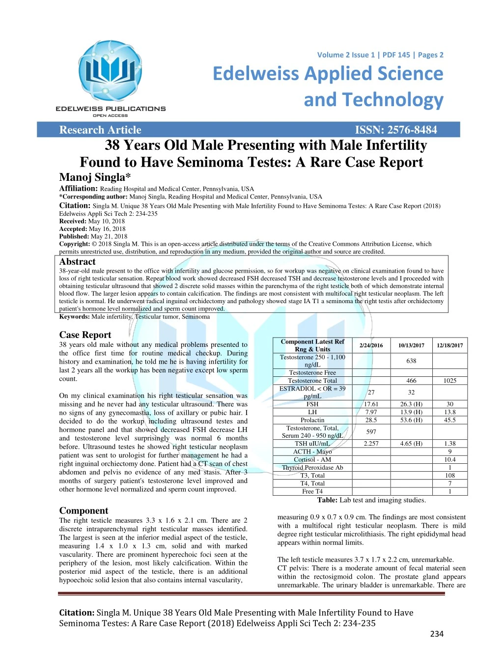
Volume 2 Issue 1 | PDF 145 | Pages 2 Volume 1 . Issue 1 | PDF 101 | Page 1 of x Edelweiss Applied Science and Technology Research Article 38 Years Old Male Presenting with Male Infertility Found to Have Seminoma Testes: A Rare Case Report Manoj Singla* Affiliation: Reading Hospital and Medical Center, Pennsylvania, USA *Corresponding author: Manoj Singla, Reading Hospital and Medical Center, Pennsylvania, USA Citation: Singla M. Unique 38 Years Old Male Presenting with Male Infertility Found to Have Seminoma Testes: A Rare Case Report (2018) Edelweiss Appli Sci Tech 2: 234-235 Received: May 10, 2018 Accepted: May 16, 2018 Published: May 21, 2018 Copyright: © 2018 Singla M. This is an open-access article distributed under the terms of the Creative Commons Attribution License, which permits unrestricted use, distribution, and reproduction in any medium, provided the original author and source are credited. Abstract 38-year-old male present to the office with infertility and glucose permission, so for workup was negative on clinical examination found to have loss of right testicular sensation. Repeat blood work showed decreased FSH decreased TSH and decrease testosterone levels and I proceeded with obtaining testicular ultrasound that showed 2 discrete solid masses within the parenchyma of the right testicle both of which demonstrate internal blood flow. The larger lesion appears to contain calcification. The findings are most consistent with multifocal right testicular neoplasm. The left testicle is normal. He underwent radical inguinal orchidectomy and pathology showed stage IA T1 a seminoma the right testis after orchidectomy patient's hormone level normalized and sperm count improved. Keywords: Male infertility, Testicular tumor, Seminoma Case Report 38 years old male without any medical problems presented to the office first time for routine medical checkup. During history and examination, he told me he is having infertility for last 2 years all the workup has been negative except low sperm count. On my clinical examination his right testicular sensation was missing and he never had any testicular ultrasound. There was no signs of any gynecomastia, loss of axillary or pubic hair. I decided to do the workup including ultrasound testes and hormone panel and that showed decreased FSH decrease LH and testosterone level surprisingly was normal 6 months before. Ultrasound testes he showed right testicular neoplasm patient was sent to urologist for further management he had a right inguinal orchiectomy done. Patient had a CT scan of chest abdomen and pelvis no evidence of any med stasis. After 3 months of surgery patient's testosterone level improved and other hormone level normalized and sperm count improved. Component The right testicle measures 3.3 x 1.6 x 2.1 cm. There are 2 discrete intraparenchymal right testicular masses identified. The largest is seen at the inferior medial aspect of the testicle, measuring 1.4 x 1.0 x 1.3 cm, solid and with marked vascularity. There are prominent hyperechoic foci seen at the periphery of the lesion, most likely calcification. Within the posterior mid aspect of the testicle, there is an additional hypoechoic solid lesion that also contains internal vascularity, ISSN: 2576-8484 Component Latest Ref Rng & Units Testosterone 250 - 1,100 ng/dL Testosterone Free Testosterone Total ESTRADIOL < OR = 39 pg/mL FSH LH Prolactin Testosterone, Total, Serum 240 - 950 ng/dL TSH uIU/mL ACTH - Mayo Cortisol - AM Thyroid Peroxidase Ab T3, Total T4, Total Free T4 2/24/2016 10/13/2017 12/18/2017 638 466 1025 27 32 17.61 7.97 28.5 26.3 (H) 13.9 (H) 53.6 (H) 30 13.8 45.5 597 2.257 4.65 (H) 1.38 9 10.4 1 108 7 1 Table: Lab test and imaging studies. measuring 0.9 x 0.7 x 0.9 cm. The findings are most consistent with a multifocal right testicular neoplasm. There is mild degree right testicular microlithiasis. The right epididymal head appears within normal limits. The left testicle measures 3.7 x 1.7 x 2.2 cm, unremarkable. CT pelvis: There is a moderate amount of fecal material seen within the rectosigmoid colon. The prostate gland appears unremarkable. The urinary bladder is unremarkable. There are Citation: Singla M. Unique 38 Years Old Male Presenting with Male Infertility Found to Have Seminoma Testes: A Rare Case Report (2018) Edelweiss Appli Sci Tech 2: 234-235 234
Singla M. Edelweiss Applied Science and Technology, 2018 PDF: 145, 2:1 normal size inguinal lymph nodes. There is no discrete adenopathy seen. There is no ascites. The appendix appears unremarkable. Impression 1. There is no adenopathy seen. 2. There is a moderate amount of fecal maternal seen in the rectosigmoid colon. Discussion Testicular cancer is the most common solid malignancy affecting males between the ages of 15 and 35, although it accounts for only 1 percent of all cancers in men [1]. Germ cell tumors (GCTs) account for 95 percent of testicular cancers . They may consist of one predominant histologic pattern or represent a mix of multiple histologic types. For treatment purposes, two broad categories of testis tumors are recognized: pure seminoma (no nonseminomatous elements present) and all others, which together are termed nonseminomatous germ cell tumors (NSGCTs). In most series, the ratio of seminoma to NSGCT is approximately one. Testicular cancer has become one of the most curable of solid neoplasms because of remarkable treatment advances beginning in the late 1970s. Prior to that time, testicular cancer accounted for 11 percent of all cancer deaths in men between the ages of 25 and 34, and the five-year survival rate was 64 percent [2]. In 2017, approximately 400 deaths from testicular cancer are expected in the United States, with a five-year survival rate of over 95 percent [1]. Testicular tumors usually present as a nodule or painless swelling of one testicle, which may be noted incidentally by the patient or by his sexual partner [3]. Occasionally, a man with a previously small atrophic testis will note enlargement. Approximately 30 to 40 percent of patients complain of a dull ache or heavy sensation in the lower abdomen, perianal area, or scrotum, while acute pain is the presenting symptom in 10 percent. In any man with a solid, firm mass within the testis, testicular cancer must be the considered diagnosis until proven otherwise. Prompt diagnosis and treatment of testicular cancer provides the best opportunity for cure. Nevertheless, both patient and clinician factors often contribute to a delay in diagnosis. The widespread use of scrotal ultrasound in the evaluation of male infertility occasionally leads to the diagnosis of an incidental nonpalpable testicular mass [4,5]. Conclusion In male infertility, physical examination is key and ultrasound of testes should be a routine part of workup. References 1.Siegel RL, Miller KD, Jemal A. Cancer statistics, 2018 (2018) CA Cancer J Clin 68:7. 2.Einhorn LH. Treatment of testicular cancer: a new and improved model (1990) J Clin Oncol 8: 1777. 3.Bosl GJ, Motzer RJ. Testicular germ-cell cancer (1997) N Engl J Med 337: 242 4.Toren PJ, Roberts M, Lecker I, et al. Small incidentally discovered testicular masses in infertile men--is active surveillance the new standard of care? (2010) J Urol 183: 1373. 5.Eifler JB Jr, King P, Schlegel PN. Incidental testicular lesions found during infertility evaluation are usually benign and may be managed conservatively (2008) J Urol 180: 261. Citation: Singla M. Unique 38 Years Old Male Presenting with Male Infertility Found to Have Seminoma Testes: A Rare Case Report (2018) Edelweiss Appli Sci Tech 2: 234-235 235