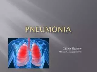
Pneumonia
Pneumonia. By: Nicole Smidt & Sarah Nainggolan. Types of Pneumonia. 1. Community-acquired (CAP) 2. Hospital-acquired (nosocomial) (HAP) 3. Ventilator-associated (VAP). (CAP) Pathophysiology. -CAP is acquired by the body via an inhalational or aspirational route.

Pneumonia
E N D
Presentation Transcript
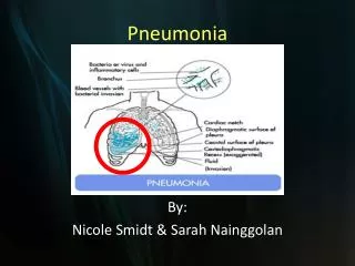
Pneumonia By: Nicole Smidt & Sarah Nainggolan
Types of Pneumonia 1. Community-acquired (CAP) 2. Hospital-acquired (nosocomial) (HAP) 3. Ventilator-associated (VAP)
(CAP) Pathophysiology -CAP is acquired by the body via an inhalational or aspirational route. -Sometimes, CAP can be obtained as a secondary bacteremia from another part of the body resulting in pneumonia caused by bacteria common to other areas of the body. -CAP tends to affect the elderly, immunocompromised, and those with underlying lung conditions (i.e. COPD, chronic bronchitis).
H&P in a (CAP) patient • History • Varying degrees of fever with acute onset • Productive cough with yellow/green sputum • Pleuritic chest pain • SOB • Physical • Auscultation of rales over the involved lobe or segment • Increased tactile vocal fremitus, bronchial breathing, and egophony (e>a sound) • Purulent sputum • Blood-tinged sputum • Signs of consolidation
Bacterial Pathogens of (CAP) ~85% of CAPs • Streptococcus pneumoniae • MCC • H. influenzae - COPD • Moraxella catarrhalis • MC in patients with chronic bronchitis & COPD
Common Pathogens/Atypical Hosts -Staph aureus causes CAP in the setting of postviral influenza. -Klebsiella pneumoniae causes CAP in chronic alcoholics. Aspiration pneumonia is typically caused by multiple pathogens. -Pseudomonas aeruginosa is a cause of CAP in patients with cystic fibrosis and bronchiectasis.
Atypical Pathogens of CAP • Zoonotic • Chlamydia psittaci • Psittacosis • Recent close contact with birds • Francisella tularensis • Tularemia • Contact with deer or rabbits or recent bite by a tick or deer fly • Coxiella burnetii • Q fever • Contact with a cat or sheep that has given birth recently • Non-zoonotic (~15% of CAPs) • Legionella • Elderly, smokers, immunocompromised • Mycoplasma pneumoniae • Young and healthy • Chlamydia pneumoniae • Young & healthy
Atypical CAP -Atypical pneumonias commonly present with dry cough, extrapulmonary involvement, and patchy interstitial pattern on CXR.
Differential Dx of CAP -Chronic bronchitis -Myocardial infarction -Asthma -CHF -Pulmonary edema -PE -Acute hypersensitivity reaction -Bronchogenic carcinoma
Laboratory Studies of CAP -CBC with differential showing a neutrophilia w/left shift, electrolytes, elevated BUN/Cr, elevated glucose, abnormal LFTs -Sputum Gram stain and/or culture (should have <10 squamous cells/lpf/purulent should have >25 PMNs/lpf) -2-3 sets of blood cultures (before antibiotics) to detect presence of bacteria in the blood -Serum cold agglutinin titers (Mycoplasma) -Urinalysis -Serological testing if zoonotic atypical pathogens are suspected
Imaging Studies of CAP -Chest x-ray *Repeat chest x-ray within 24 hours if first x-ray shows negative findings with high clinical suspicion -Serial chest x-rays to observe for progression or check for resolution -Chest CT scan *If failing to respond to therapy
Community Acquired Pneumonia (CAP) Treatment • Decision to hospitalize • Pneumonia Severity Index (PSI) • Points given for 20 variables i.e. age, comorbid conditions, abnormal PE & lab findings • Add up the points; patients are categorized into 5 classes
Decision to hospitalize, cont. 2. CURB-65 • Five variables • C=Confusion • U=Urea > 7mmol/L • R=Respiratory rate ≥ 30 • B=BP systolic ≤ 90, diastolic ≤ 60 • Age ≥ 65 • Add up the points, patients are categorized into 3 classes
Decision to hospitalize, cont. • CURB-65, cont. • PSI-less practical • CURB-65-less studies done
CAP Treatment • Empiric antibiotic treatment • Outpatient treatment • Previously healthy or no antibiotics in last 3 months • Comorbidities or antibiotics in last 3 months • Inpatient treatment • Non-ICU patients • ICU patients
CAP TreatmentOutpatient antibiotics Guidelines per IDSA and ATS
CAP Treatment Special Considerations • Pseudomonas • piperacillin/tazobactam 4.5 g IV q6h, cefepime 1–2 g IV q12h, imipenem 500 mg IV q6h, meropenem 1 g IV q8h plus either ciprofloxacin 400 mg IV q12h or levofloxacin 750 mg IV, or • piperacillin/tazobactam 4.5 g IV q6h plus amikacin 15 mg/kg qd or tobramycin 1.7 mg/kg qd and azithromycin, or • piperacillin/tazobactam 4.5 g IV q6h plus an aminoglycoside plus an antipneumococcal fluoroquinolone • If PCN-allergic: substitute aztreonam
CAP TreatmentSpecial Considerations • Community-Acquired MRSA • Add linezolid 600 mg IV q12h or vancomycin 1 g IV q12h
CAP Treatment • Most patients admitted to the hospital for CAP receive IV antibiotics • A switch to oral drugs is appropriate, so long as the patient can ingest and absorb the drugs, is hemodynamically stable, and showing clinical improvement • Duration: Patients are usually treated for 10-14 days, but recent studies with fluoroquinolones suggest that a 5-day course is sufficient for uncomplicated CAP.
CAPFollow-up • Fever usually resolves within 2 days, while leukocytosis may improve within 4 days • Abnormalities in CXR take 4-12 weeks to clear, depending on the age of the patient and the underlying lung disease • Follow-up CXR can be done 4-6 weeks later • If recurrence is seen in the same lung segment, possible neoplasm should be taken into consideration
CAPPrognosis • Depends on age, comorbidities, inpatient/outpatient • Young patients without comorbidities fully recover in 2 weeks • Older patients with comorbidities take longer • Overall mortality rates • Outpatient: < 1% • Inpatient: 10%, with about 50% of the deaths directly attributable to pneumonia
Healthcare-associated Pneumonia 1. Hospital-acquired pneumonia (HAP) 2. Ventilator-associated pneumonia (VAP) The main difference from CAP: significantly lower incidence of atypical pathogens, EXCEPT Legionella
Hospital-acquired pneumonia (HAP) Defined as pneumonia that occurs more than 48 hours after admission but that wasNOT incubating at the time of admission. The American Thoracic Society further subdivides nosocomial pneumonia into early (occurring within the first 4 days of hospitalization) and late (occurring after the 5th day of admission) onset.
HAP Pathophysiology -HAP is primarily acquired though aspiration of oropharyngeal secretions into the trachea. -Although uncommon, hematogenous spread is also possible. -HAP is mainly caused by aerobic gram-negative bacilli of two forms: necrotizing and nonnecrotizing. *Necrotizing cause rapid cavitation, microabscess formation, blood-vessel invasion, and hemorrhage (i.e. P. aeruginosa).
HAP -Usually bacterial in nature. -Common in patients with mechanical ventilation as intubation and ventilation support bypass the normal host defense mechanisms. -One of the most common diagnoses in the medical and surgical ICUs.
H&P of HAP History: -SOB -Productive cough -Fever Physical: -Rales on auscultation over affected area -Generally NO consolidation in nosocomial pneumonia
HAP Microbiology -P. aeruginosa -Klebsiella -E. coli -Acinetobacter (common in ICU patients) -Staph aureus -Strep pneumoniae -H. influenzae
Dx of HAP Laboratory: -White blood cell count -Blood cultures Imaging Studies: -Serial chest x-rays to check for progression and resolution -CT or spiral CT scan
Hospital-acquired Pneumonia (HAP) • Main differences from VAP: • Lower incidence of MDR pathogens (allows more monotherapy) • Better host immunity • Anaerobes are more common • Specific therapy aimed at anaerobes is not indicated, unless aspiration is a concern • Lower risk of antibiotic failure in comparison to VAP • Lower mortality rates than VAP
Ventilator-associated pneumonia Defined as pneumonia that develops 48 hours or longer after mechanical ventilation is given via endotracheal tube or tracheostomy.
VAP Pathophysiology -VAP is caused by invasion of the lower respiratory tract and lung tissue by microorganisms caused by decreased integrity of the oropharynx and trachea allowing oral and gastric sections to enter. -The incidence of VAP increases with the duration of ventilation. -Pseudomonas and Acinetobacter pneumonia is associated with the greatest mortality.
Dx of VAP History: 1. Hospitalization of greater than 5 days 2. Hospital admission of more than 2 days in the last 90 days 3. Antibiotic use in the last 90 days 4. Residing in a nursing home or skilled care facility 5. Home infusion therapy or wound care 6. Dialysis within the last 30 days 7. Immunocompromised Diagnostic Triad: 1. Fever 2. Purulent secretions 3. Leukocytosis
Work-up of VAP -CBC with differential to evaluate white blood cell count -Routine blood tests to assess patient's baseline renal and hepatic function to properly dose antibiotics -Blood cultures -Respiratory secretion cultures -Portable chest radiography -Air bronchogram
Ventilator-associated Pneumonia (VAP) • Empirical Antibiotic Treatment
VAP Treatment • Duration: 8 days (associated with less frequent emergence of antibiotic-resistant strains) • Pseudomonas aeruginosa: causes high rates of clinical failure and death, despite combo therapy • VAP caused by MRSA: 40% clinical failure rate when treated with standard-dose vancomycin, so treat with linezolid instead.
VAPFollow-up • Clinical improvement is usually seen within 48-72 hours following initial antibiotic therapy • CXR worsens during initial treatment, so less helpful • In very ill patients, follow-up CXR can be done every few days • Once patient has improved, CXR may not be necessary for a few weeks
VAPPrognosis • Associated with high crude mortality rates (50-70%) • Stenotrophomonas maltophilia: prognosis is very poor (death is almost inevitable)
Question #1 From USMLEWORLD • A 65-year-old white male who has a history of COPD, CHF, and CAD presented with a three-day history of worsening SOB, cough with yellowish expectoration, and fever. He is not on steroids and does not use oxygen at home. He takes ipratropium, albuterol, aspirin, digoxin, furosemide, metoprolol, and lisinopril. He lives at home with his wife. His temp is 103 F, BP is 110/70, pulse is 110, and respirations are 24. He is saturating 88% on room air. He was started on 3-liters of oxygen to keep the saturations above 92%. The CXR of the patient is shown below. What is the most appropriate next step in the management of this patient? A. Admit the patient and give ampicillin B. Outpatient trimethoprim-sulfamethoxazole C. Admit the patient and start levofloxacin D. Admit the patient and start ciprofloxacin E. Admit the patient and start vancomycin F. Outpatient clindamycin therapy
Question #1 CXR
Answer #1 • C. Admit the patient and start levofloxacin • The clinical presentation and CXR of this patient is highly suggestive of CAP. The first step in the management of CAP is to decide whether the patient needs hospitalization. The decision to admit the patient is generally made using the Pneumonia Severity Index (PSI) based on medical history, PE, and lab and radiographic findings. This patient has other comorbidities such as CHF, COPD, and CAD, and his oxygen saturation is less than 90%. Thus he falls under risk class III or IV and needs to be hospitalized. • The most common cause of lobar pneumonia in this setting is pneumococcus. The new generation flouroquinolones (i.e. levofloxacin) is used for the inpatient treatment of CAP. About 97% of the Streptococcus pneumonia are sensitive to these drugs. They also cover the atypical organisms. • For the management of outpatient therapy in stable patients with no comorbidities, either azithromycin or doxycycline can be used.
Question #2 A 68-year-old woman with polymyositis is evaluated in the office because of increased difficulty swallowing and a 2-week history of low-grade fever, intermittent cough, and sputum production. On physical examination, temperature is 37.9 °C (100.2 °F), and crackles are heard at the base of the right lung posteriorly. The leukocyte count is 9700/μL with 85% neutrophils, 12% lymphocytes, 2% monocytes, and 1% eosinophils. A chest radiograph shows patchy pulmonary infiltrates in the right lower lobe. Sputum Gram stain shows many neutrophils, a few squamous epithelial cells, and several morphologic types of both gram-negative rods and gram-positive cocci. Results of sputum culture are pending. Which of the following is the most appropriate management at this time? A. Bronchoscopy B. Clindamycin C. Metronidazole D. Penicillin
Answer #2 B. This patient should be started on clindamycin. The presence of underlying polymyositis and difficulty swallowing place this patient at risk for aspiration and the development of aspiration pneumonia. Other risk factors for aspiration pneumonia include episodes of depressed consciousness (e.g., patients with alcoholism) or mechanical factors that increase the likelihood of secretions entering the tracheobronchial tree (e.g., patients with esophageal obstruction). Although aspiration of acidic gastric contents can result in a chemical pneumonitis, aspiration of oral secretions typically results in an anaerobic pneumonia caused by anaerobic organisms that are normally present in the mouth. A sputum Gram stain often shows both gram-positive and gram-negative organisms of different morphologic types, as was found in this patient. The typical location of aspiration pneumonia depends upon the patient's position at the time of aspiration. Based on gravitational flow of airway secretions, the pneumonia is preferentially localized in the dependent regions of the lung at the time of aspiration. In an upright patient, the lower lobes are usually affected, more so on the right than on the left. In a supine patient, common sites of aspiration pneumonia are the posterior segment of the right upper lobe and the superior segment of the right lower lobe. Although options for antibiotic coverage of anaerobic aspiration pneumonia have generally included either penicillin or clindamycin, controlled studies have shown that antibiotic failure rates have been higher for penicillin than for clindamycin. Metronidazole does not adequately cover microaerophilic and aerobic streptococci from the mouth, which are contributing organisms in many patients with aspiration pneumonia. Bronchoscopy is not indicated because it is unable to confirm anaerobic infection, since anaerobic organisms from the mouth will contaminate the specimens obtained.
References • Mandell LA, Wunderick R, et al. Harrison’s Principles of Internal Medicine, 17th ed. 2008. • Chesnutt MS, Gifford AH, Prendergast TJ. Current Medical Diagnosis & Treatment 2010. • Agabegi SS, Agabegi ED. Step-Up to Medicine, 2nd ed. 2008. • Fischer C, Reichert S. USMLE Master the Boards. 2009. • Burke A Cunha, MD. Emedicine.medscape.com. 2010.