
Impacts of Dasatinib on Phospho-Src and c-Kit in Breast Cancer Cell Lines
This study investigates the effects of dasatinib on the phospho-Src and c-Kit levels in various breast cancer cell lines, including MDA-MB-468, MDA-231, and SKBR-3. Whole cell lysates were obtained from untreated cells and subjected to Western blotting to determine basal levels of phospho-Src and c-Kit, with GAPDH as a loading control. MDA-MB-468 cells treated with dasatinib for 48 hours were assessed using an MTT assay. Results indicated dosages, with an IC50 of 17.8 μM for dasatinib, showcasing treatment efficacy in reducing cell viability.

Impacts of Dasatinib on Phospho-Src and c-Kit in Breast Cancer Cell Lines
E N D
Presentation Transcript
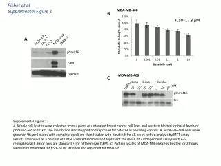
Pichot et alSupplemental Figure 1 B IC50=17.8 μM MDA-231 MDA-468 SKBR-3 T47D A MDA-MB-468 MCF-7 Dasa Doxo Combo (nM) DMSO 100 100 100 10 10 10 pSrc416 1 pSrc-Y416 Src c-kit GAPDH C Supplemental Figure 1: A, Whole cell lysates were collected from a panel of untreated breast cancer cell lines and western blotted for basal levels of phospho-Src and c-kit. The membrane was stripped and reprobed for GAPDH as a loading control. B, MDA-MB-468 cells were grown in 96-well plates with complete medium, then treated with dasatinib for 48 hours before analysis by MTT assay. Results are shown as a percent of DMSO-treated samples and represent the mean of 2 independent assays with 4-5 replicates each. Error bars are standard error of the mean (SEM). C, Protein lysates of MDA-MB-468 cells treated for 2 hours were immunoblotted for pSrc-Y416, stripped and reprobed for total Src.