Download
1 / 30
780 likes | 3.98k Views
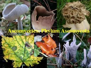
Division Ascomycota. Somatic stage Hyphae or yeast form Septate hyphae with simple septum and woronin bodies are presence Often organize into fungal tissue, e.g. stroma and sclerotia Cell wall contains chitin mainly Spindle pole body Reproduction

E N D
Somatic stage Hyphae or yeast form Septate hyphae with simple septum and woronin bodies are presence Often organize into fungal tissue, e.g. stroma and sclerotia Cell wall contains chitin mainly Spindle pole body Reproduction Asexual reproduction by production of conidiospore, chlamydospore, and fragmentation. Fission and budding are performed by yeast. Sexual reproduction by formation of “ascospores” within a sac-like cell termed “ascus” u u Main characteristics
Sexual reproduction: Ascospore formation A: ascogonium + antheridium; B: plasmogamy; C: dikaryotic mycelium; D: crozier hook; E-F: nuclear division; G: karyogamy; H: meiosis; I: mitosis; J: ascus with ascospores
Sexual reproduction: Ascus Usually elongated cylindrical, but can be globous or spherical form Presence or absence Presence or absence 1. Protunicate 2. Unitunicate 3. Bitunicate Shape: Stalk: Operculum: Asci structure:
Sexual reproduction: Ascus structure Protunicateasci are usually more or less spherical and are found in cleistothecia. They have a thin, delicate wall and release spore by deliquescing due to they have no spore-shooting mechanism. Unitunicate asci have a single wall (endotunica and exotunica are fused) thoughout the existence of the ascus. The spores are released through a terminal pore, slit or operculum. Bitunicate asci have double wall, i.e. endotu- nica and exotunica. The outer wall is thin and inextensible, while the inner wall is thick and elastic. Prior to spore release, the endotunicata usually expands and protrudes from the ruptured exotunica and spores are release through a pore of endotunica.
Sexual reproduction: Ascocarp Ascocarp is a fruiting body bearing asci.According to ascocarp structure, there are 5 types can be distinguished. 1. Naked asci (no ascocarp) 2. Cleistothecium 3. Perithecium 4. Apothecium 5. Pseudothecium or ascostroma
Taphrina deformans Eremascus albus Sexual reproduction: Naked asci The fungi form asci directly from zygotes or single cells, without ascogenous hyphae or ascocarp. Saccharomyces cerevisiae
Sexual reproduction: Cleistothecium Cleistothecium is a completely closed ascocarp and have asci that are scattered throughout the interior that is to call without hymenium. Break- down of the cleistothecium might be only one way to release the ascospore
Sexual reproduction: Perithecium Perithecium is various shaped, but typically flask-shaped or globous with a small ostriole. Asci are arranged in a single fertile layer throughout the base of perithecium and this fertile layer is called “hymenium”. Paraphyses, sterile cells, may be present among the asci.
Jafneadelphus ferrugineus Sexual reproduction: Apothecium Aphthecium is usually cup-shaped or variable. The asci form hymenium that entirely exposed. Paraphyses are generally present in apothecia
Sarcoscypha coccinea Sarcoscypha mesocyatha Leotia lubrica Gyromitra californica Morchella esculenta Sexual reproduction: Apothecium
Sexual reproduction: Ascostroma (pseudothecium) Ascostroma is a locule that forms in a stroma where the asci are borne. This differs from a perithecium that is formed within a stroma in that a perithecial wall is formed by the perithecium that delimits it from the stroma. Elsinoe leaf spot Elsinoe vacinii
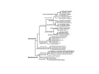
Classification: The new classification New classification of Ascomycota based on molecular information (Sugiyama, 1993; Sukiyama and Nishida, 1994) Class: Euascomycetes (Filamentous ascomycetes) Class: Protoascomycetes (Yeast and Archiascomycetes) Class: Laboulbeniomycetes
Ascocarp Group/ Class/ Series Non-ascocarp Cleistothecium Perithecium Apothecium Ascostroma Hemiascomycetes Plectomycetes Pyrenomycetes Discomycetes Loculoascomycetes Laboulbeniomycetes Hymenoascomycetes Classification: The classification in our class A very old simply and classic classification (modify from Alexopolos and Mims, 1979)
Hemiascomycetes: Order Protomycetales (yeast) Species Saccharomyces cerevisiae Schizosaccharomyces pombe Candida utilis Candida albicans Cryptococus neoformans Pneumocystis carinii Importance Fermentation, brewery, molecular model Molecular model Food yeast Superficial and systemic infection Pulmonary disease Effect to central nervous system Pneumonia
Naked asci of Taphrina deformans peach and leaf curl Hemiascomycetes: Order Taphrinales Order Taphrinales are all parasite on plant. A well know species in this order is Taphrina deformans, peach and almond leaf curl agent.
Plectomycetes: Chalkbrood due to Ascosphera apis Top-left: Infected larvae in their cells Top-right: Mummify larvae Bottom: Cleistothecia containing asci of Ascosphera apis
Plectomycetes: Powdery mildew (ราแป้ง) Poedwey mildew is a plant disease caused by Order Erysiphales. Some serious pathogens are Uncinulla necator, Erysiphe graminis and Sphaerotheca spp.
Plectomycetes: Dermatophytes (ราก่อโรคผิวหนัง) โรคกลาก (Tinea หรือ Ringworm) มีสาเหตุมาจากเชื้อราชนิดที่เป็น dermatophyte คือเชื้อราที่สามารถย่อย keratin ได้ เชื้อโรคกลาก อาจพบได้ในรา 3 สกุล คือ genus Epidermophyton, Microsporum และ Trichophyton ซึ่งมีลักษณะของ asexual spores ที่แตกต่างกัน Microsporum Trichophyton Epidermophyton
Plectomycetes: Dermatophytes (ราก่อโรคผิวหนัง)
Pyrenomycetes: Flask fungi Hypoxylon sp. Xylaria sp. Xylaria sp. Xylaria sp. Perithecium
Pyrenomycetes: Ergot of rye The sclerotia of Claviceps purpurea, the cause of ergot of rye, produce a number of powerful alkoloid, for example ergotamin, ergometrin and ergonovin, which are used as medicine to induce labor and prevent post partum hemorrhage during childbirth.
Pyrenomycetes: Cordyceps Cordyceps spp., the member of F. Clavicipi- taceae are parasitic on either insect or hypo- genous ascocarp. Cordyceps are also used as traditional chainess medicine.
Discomycetes: Cup fungi Vibrissea sp. Piziza sp. Cookeina sp. Scutellinia sp. Microstroma sp.
Discomycetes: Morel Verpa sp. Morchella esculenta
Loculoascomycetes Loculoascomycetes are generally characterized by the production of bitunicate asci within stromatic locules. Ascocarp of Venturia inaequalis Apple scab