Download
1 / 37
370 likes | 464 Views
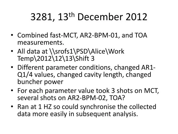
3281, 13 th December 2012. Combined fast-MCT, AR2-BPM-01, and TOA measurements. All data at \srofs1PSDAliceWork Temp20121213Shift 3 Different parameter conditions, changed AR1-Q1/4 values, changed cavity length, changed buncher power

E N D
3281, 13th December 2012 • Combined fast-MCT, AR2-BPM-01, and TOA measurements. • All data at \\srofs1\PSD\Alice\Work Temp\2012\12\13\Shift 3 • Different parameter conditions, changed AR1-Q1/4 values, changed cavity length, changed buncher power • For each parameter value took 3 shots on MCT, several shots on AR2-BPM-02, TOA? • Ran at 1 HZ so could synchronise the collected data more easily in subsequent analysis.
Summary 5nd Feb • FEL pulse energy varies (within the shot) from 6% to 14% rms on the subset of data I had looked at. • FEL pulse frequency spectrum • ~100 KHz always there. ‘Two-spike’ structure as seen in BPM data • 300 KHz sometimes observable, sometimes not. • High frequency content 1-8 MHz, in DFT and can see regular high frequency by eye. Insufficient sampling of the FEL pulses? Or real effect? Use Genesis to investigate? • 800 kHz - and seen in BPM?
New Updates February • 5 MHz • Can’t be aliasing effect based on pulse freq and sampling freq (AK) • Does it depend on buncher phase ? No see slide 12 • Other lower high frequency components (~ 3 MHz) could be due to aliasing • Genesis simulations
Examples of MCT signal and Fourier analysis • Took the earliest time labelled MCT data • \\\\Srofs1\\psd\\Alice\\Work Temp\\2012\\12\\13\\Shift 3\\timingRawData\\C2-214519-632-A00000.trc • 3 shots in each file, look at first shot • Sampling is 410-10 sec, or 2.5 GHz individual pulses are resolved
Frequency Content … of example shot detailed on previous slide FULL DFT Max frequency = 2.5/2 GHz = 1.25 GHz ~ 70 μsec section of pulse train MHz 16 MHz bunch rep rate + harmonics Familiar frequency profile with 100 KHz and 300 kHz, seen in BPM DFTs MHz MHz
MCT Frequency Content • I think the MCT variations, at the same frequency as what is observed on the BPM position, makes sense. • FEL build-up/decay time is on the order ~ 1 μsec. So one FEL pulse is influenced by ~ 1 μs of bunches. If the next ~ 1 μsec set of bunches is offset with respect to the previous set, they will produce pulses of a different intensity (since beam offset influences FEL intensity strongly). • Thus the FEL pulse intensity should exhibit some sort of moving average of the bunch positions, where the moving average is over ~ 1 μs. Thus bunch position variations on a 10 μs (100 kHz) scale would not be smeared out. • Bunch charge variations of 300 kHz (~ 3 μs) might also be visible.
Analysis Issues • My initial look at the data, on previous slides, computed MCT_i - <MCT> where i = the ith MCT voltage measurement then took the DFT • Probably the wrong thing to do, since MCT_i contains all the MCT noise around between FEL pulses as well as the FEL pulses • Really, only want the peak MCT value from each FEL pulse • But scope sampling is not fast enough to catch the maximum MCT peak value with high accuracy, see next slide • This was not quite the case with PI laser pulse power in previous shifts (e.g. #3205) since sampling rate was sufficient in that case.
Analysis Issues • Rising edge of measured FEL pulses are not caught by the 0.4 ns sampling spacing. • This may introduce an additional artificial variation in measured pulse-to-pulse FEL power if only the peak values are taken • But if DFT is done on complete data, the variation of ALL the measurement points is computed and perhaps the physical variations of FEL pulse power should become larger relative to the unphysical variation due to finite sampling effects. • However, measuring things like rms FEL pulse energy variation might be difficult to subtract this spurious variation due to sampling. (Integrate pulses to reduce effect)
Analysis Issues • Integrate each peak to get pulse by pulse MCT energy • Not as easy as you might think. It’s not actually easy to find, count and integrate the peaks, even for these very regularly spaced peaks, even with only little noise between them. • Solution found at http://forums.wolfram.com/mathgroup/archive/2009/Dec/msg00364.html
General Frequency Observations • MCT shots taken over many different machine parameters (~ 50 different conditions, cavity length, quad strength, buncher power) • Most show frequency content around ~ 100 kHZ, sometimes a single peak, sometimes two peaks, as seen in BPM data • Higher frequency components visible 1-8 MHz. • 300 kHz sometimes there but amplitude is not as pronounced, and often it’s not discernible (got lucky seeing it on the first example slides 1-2). On BPM data, and INJ-FCUP, PI laser, #3205 300 kHz signal was always discernible. • Other frequencies (e.g. 800 kHz) depending on data processing of MCT signal.
General High Frequency Observations. Examples, varying buncher phase FEL pulse integrated, normalised to mean value, then perform DFT -> the DFT values are fractional FEL power variation Buncher phase not backlash corrected φ= 256 φ= 254 High frequency 0-8 MHz φ= 262 φ= 264 φ= 260 φ= 258 MCT pulse peak value vs pulse number The high frequency spikes are there and don’t change much, whatever MCT data is used : complete, peak value, integrated pulse
Replot High Freq, Buncher Phase Vary High Freq DFT of integrated FEL pulse energy Not an obvious dependence of the ~ 5 MHz component on buncher phase If you take peak MCT value per pulse, rather than integrated, the 5 MHz spikes are not affected drastically but other high frequency components appear. These are the DFT of the first of three shots for each buncher phase. The 2nd and 3rd show very similar spectra (same frequency components, same amplitude)
General Low Frequency Observations. Examples, varying buncher phase FEL pulse integrated, normalised to mean value, then perform DFT -> the DFT values are fractional FEL power variation Low frequency 0-1 MHz ALL MCT data taken, normalised to mean value, then perform DFT -> the DFT values are fractional FEL power variation -> Note high frequency components In several of these spectra you might not ‘see’ the 300 kHz if you weren’t specifically looking for it. In fact the more obvious content is at ~ 800 kHZ The DFT amplitude of variation of the ~ 100 KHz component is up to 12%
BPM Observations • To synchronise BPM shot with MCT shot use time stamps. • Use first example MCT data at \\Srofs1\psd\Alice\Work Temp\2012\12\13\Shift 3\timingRawData\C2-223058-304-mirror-308.7A00000.trc as it has quite prominent 800 kHz and ~ 5 MHz components • e.g. MCT time stamp TRIGGER_TIME,{35.8057,2.,22.,13.,12.,2012.,0.} • 35.8 seconds after 2202 on 13th. • Take file FELbase_Buncher260_220235-195_BPM_08.bpm • Each shot is time-stamped, take shot 2012-12-13 22:02:35.993630 as it’s the closest Don’t think this is correct way of synchronising
BPM Observations – x FELbase_Buncher260_220235-195_BPM_08.bpm 2012-12-13 22:02:35.993630 No sign of 5 MHz DFT MCT (pulse integrated) 800kHz
BPM Observations – y FELbase_Buncher260_220235-195_BPM_08.bpm 2012-12-13 22:02:35.993630 No sign of 5 MHz DFT MCT (pulse integrated)
5 MHz? (+ other components) all data MCT peak value vs pulse number pulse maximum BPM position pulse integrated peak value high frequency component visible in the data DFT spectrum vs MCT treatment method
5 MHz? ( + other components) What causes them? Sampling artefact in the MCT data ? (see slide 5) i.e. Aliasing effect ? Seems plausible, but the amplitude of the ~ 5 MHz spike doesn’t change however you treat the data (full data, peak value, integrated peak). If it is an aliasing effect wouldn’t you expect the 5 MHz amplitude to change depending on the data treatment? Or some detector MCT DAQ effect? (but the size of the ~ MHz components vary shot by shot)
MCT pulse-to-pulse variation • As seen from the DFTs in examples on slide 9, the amplitudes of the ~ 100 kHz components can be up to 12%. (DFTs normalised such that their values give the equivalent amplitudes of sine-functions with the same frequencies) • Also take RMS of slide 9, the RMS normalised FEL pulse energy varies (within the shot) from 6% to 14%
Genesis Simulations of ALICE FEL GENESIS INPUT FILE gamma0= 54.81600E+00 delgam= 0.162900E+00 rxbeam= 2.260000E-04 rybeam= 4.775000E-04 alphax= 0.000000E+00 alphay= 1.750000E+00 emitx = 1.200000E-05 emity = 1.200000E-05 • Genesis + OPC (optical propagation code), time independent. • Genesis simulates the FEL process along a single pass of the undulator, OPC propagates the radiation with mirrors. • Don’t exactly know how it all works yet. • Can vary the position of the beam at each pass (pass == bunch). • Test the effect of bunch position variations on FEL pulse intensity low frequency • Information on possible causing high frequency components? Need time-dependent simulation
Genesis ALICE FEL simulations FEL pulse power vs pulse number 0.2 mm sin x variation with 0.2 mm global x offset λ = 100 bunches Default settings, no beam offset 0.2 mm sin x variation λ = 100 bunches Abs power Relative power Expected pulse power = ~ 1 μJ/1 ps 1 mW (NIM first lasing paper)
Cavity Length Scans – MCT This data is superseeded by Trina’s synchronised MCT/BPM/TOA data set • MCT of saturated part Pulse power getting very low, peak finding algorithm starts to fail These plots show MCT Max vs pulse number. DFTs operate on MCT integrated pulse voltage vs pulse number
Cavity Length Scans – MCT DFT low freq This data is superseeded by Trina’s synchronised MCT/BPM/TOA data set
Cavity Length Scans – MCT DFT hi freq This data is superseeded by Trina’s synchronised MCT/BPM/TOA data set
Cavity scan. Corresponding BPM traces • Prelim. – Did not synchronise the shots properly so did not continue with Fourier. This data is superseeded by Trina’s synchronised MCT/BPM/TOA data set
\\ \\ dlfiles03 \\ Astec \\ Projects \\ ALICE \\ Work \\ 2012\\ 12\\ 13\\ Shift 3\\ SynchronisedDataSets FROM NOW ON USE TRINA’s synchronised data set
MCT RAW AR1Q vary cavity mirror vary
TOA fourier at linac re-entry (chopped), areas, smoothed, high freq some high frequency at n*1.25 MHz
TOA fourier at linac re-entry (chopped), areas, smoothed, high freq.