Slide Note
0 likes | 13 Views
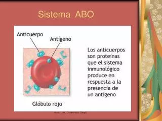
Analysis of B blood group subgroups, factors affecting antigen expression, case studies on ABO anomalies and HDFN, laboratory techniques like adsorption/elution, and maternal antibody titration to manage HDFN risks.

E N D
B Blood Group Julie Long, Senior Medical Scientist, Red Cell Immunohaematology Laboratory
B Blood Group • Ireland 11% of the population have the B blood group: • 9% B+ • 2% B-
B Subgroups Quantitative or Qualitative. • Subgroups o weak or mixed field reactions in the forward group or o discrepant reverse groups. • Important to consider other factors also: o Diseases such as acute myeloid leukaemia or other haematological cancers can lead to the depression of ABO antigens. o Medications which may cause AIHA or direct binding to red cells. o Cold reactive antibodies. o Acquired B •
B Subgroups Phenotype Anti-A Anti-B Anti- AB Anti-H Common Antibodies Unexpected Antibodies Saliva Secretors B transferase B 0 4+ 4+ 2+ Anti-A None B, H Yes B3 0 ++mf ++MF 3+ Anti-A None B, H Yes (weak) Bx 0 wk wk 3+ Anti-A Weak anti-B H No Bm 0 0/wk 0/wk 3+ Anti-A None B, H Yes (weak) Bel 0 0 0 3+ Anti-A Possible anti- B H No
Case Study 1: To B or Not to B! Automated (Ortho Vision) Tube Card (BioRad Card) 5-year-old female Myelomonocytic leukemia. Query ABO Anomaly Forward and reverse group do not match. • • Anti-A - - - Anti-B - + - / +w* • • Anti-D C C C Anti-D C C C Control - - - Reverse group: • Tube Technique 4C incubation for 1hr • Increased plasma to red cell ratio (2:1) A1 Cells V ++s ++s B Cells - - - *checked micro
Additional Laboratory Testing • C+ E- c- e+ K- • Antibody Screen: Negative • DAT results: IgG- IgM- IgA- C3d- C3c- • Auto Test cell: Negative
Adsorption / Elution technique. Add Saline Centrifuge Pooled plasma from 5 group A donors (Poly anti-B anti- sera) Anti-B is adsorbed onto the patients red cells by incubation at 4◦C for 1hr. Remove supernatant and wash red cells to remove unbound antibody. Removing of bound antibody through heat elution at 51◦C for 10 minutes.
Adsorption/ Elution technique. Test the eluate against group B and O Cells: Ads/Elution IAT RT Tube Eluate: V233866-B +s ++s +s Wash: V233866-B - - - Eluate: V233872-O - - • This technique yielded only anti-B in the eluate indicating the presence of B antigen on the red cells. Controls: • Final wash control to ensure unbound antibody is washed away and only bound antibodies are being detected. • Eluate tested against group O cells ensuring reactivity due to anti-B. • Additional control, adsorbed elution performed on a known group B sample in parallel .
Conclusion The weakened expression of the B antigen is most likely due to the patient’s clinical condition. • Diminished expression of ABH antigens can occur in hematological disorders such as leukemia, myeloproliferative disorders, myelodysplastic syndrome and in some cases of Hodgkin's lymphoma. • These modifications of blood group antigens usually revert to normal after remission is attained. •
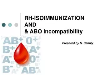
HDFN Haemolytic disease of the foetus/ new-born (HDFN) • The foetus/new-born’s red cells are being attacked by maternal IgG antibodies. A major cause of HDN is an incompatibility of the Rh blood group between the mother and fetus. • • HDN due to ABO incompatibility is usually less severe than Rh incompatibility. • The impact of HDFN ranges from mild to severe with symptoms such as edema and jaundice. •
Case Study 2: HDFN due to ABO mismatch ABO incompatibility resulting in HDFN occurs in approximately 4% of all deliveries generally causing mild haemolysis. • However, some of these cases can present as moderate or severe haemolytic disease of the foetus and new-born (HDFN) with significant hyperbilirubinemia requiring intervention. • Fetal assessment is not routinely required but early recognition based on detailed maternal clinical history and laboratory investigation is key in preventing neonatal morbidity and mortality. •
Case Study 2: • ?HDFN due to maternal anti-B. • • Mother: group O+ Baby: group B+ • The neonate required phototherapy. • Two previous pregnancy were also affected.
Laboratory Testing of Maternal Sample Group: O RhD+ Phenotype: R1R1 K- Antibody Investigation: negative Auto test cell and DAT: negative Low incidence antibody investigation: Cw, Kpa, Cob, Wra, Bga, HLA excluded. Compatibility testing: Paternal red cells versus maternal plasma and compatible result obtained .
Testing of Maternal sample continued IgG Antibody Titration • Patient plasma is incubated with neutr-AB (1:1) at 4◦C for 1hr • The neutr-AB neutralised out IgM antibodies. • A doubling dilution is prepared using both neat and neutralised patient plasma. • Direct spin and IAT tube techniques are used for the detection of IgM and IgG anti-B in the neat and neutralised dilutions
Titration Results • Titration of Anti-B in maternal plasma versus B cells using direct spin and IAT techniques. Dilutions 2 4 8 16 32 64 128 256 512 1024 IgM Anti-B Near Plasma C C C V V ++ +S - - - IgM Anti-B Neutralised - - - - - - - - - - IgG Anti-B Neautralised C C C C C V ++ + - -
Laboratory Testing on Neonatal Sample Group: B RhD+ Phenotype: R1r DAT: IgG 2+, C3d -, IgA -, IgM -, C3c – Antibody screen: Negative, but anti-A and Anti- B detected. Elution: Anti-B was detected in the eluate prepared from the neonatal red cells.
Report Conclusion Maternal Results • Antibody investigation was performed on this sample and no clinically significant antibodies were detected. • The patient's plasma was tested against the following panel of low incidence antigens and no reactivity was observed; Cw, Kpa, Lua, Wra, Cob, Kpa, Bga, HLA, Ytb. • An Immune anti-B titration was performed and IgG anti-B titrated at 256. • An Immune anti-A titration was performed and IgG anti-A titrated at 32. Neonate Results • Following three cell antibody screen, no clinically significant antibodies were detected. The patient's neat plasma was tested against A1 & B cells and reactivity was observed with both A1 & B cells. This is presumably maternal in origin. • An eluate was prepared from the patient's red cells and anti-B was detected. This is presumably maternal in origin.
References Nambiar, R. K., Narayanan, G., Prakash, N. P., & Vijayalakshmi, K. (2017). Blood group change in acute myeloid leukemia. Proceedings (Baylor University. Medical Center), 30(1), 74–75. Akanmu, A. S., Oyedeji, O. A., Adeyemo, T. A., & Ogbenna, A. A. (2015). Estimating the Risk of ABO Hemolytic Disease of the Newborn in Lagos. Journal of blood transfusion, 2015, 560738. Dean L. Blood Groups and Red Cell Antigens [Internet]. Bethesda (MD): National Center for Biotechnology Information (US); 2005. Chapter 4, Hemolytic disease of the newborn. Available from: https://www.ncbi.nlm.nih.gov/books/NBK2266/ • • •
Thank you for listening.


![[PDF] Free Download The Blood Mirror By Brent Weeks](https://cdn4.slideserve.com/8010772/slide1-dt.jpg)
![[PDF] Free Download Blood Of Iax By Robbie MacNiven](https://cdn4.slideserve.com/8018502/slide1-dt.jpg)
![[PDF] Blood Communion](https://cdn4.slideserve.com/8031337/slide1-dt.jpg)
![[PDF] Free Download Fire and Blood By George R.R. Martin & Doug Wheatley](https://cdn4.slideserve.com/8142107/slide1-dt.jpg)