Download
1 / 10
100 likes | 245 Views
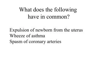
What does the following have in common?. Expulsion of newborn from the uterus Wheeze of asthma Spasm of coronary arteries. Basics of muscle contraction. Control of intracellular Ca 2+ - principal mechanism that initiates contraction and relaxation in smooth and striated muscle

E N D
What does the following have in common? Expulsion of newborn from the uterus Wheeze of asthma Spasm of coronary arteries
Basics of muscle contraction • Control of intracellular Ca2+ - principal mechanism that initiates contraction and relaxation in smooth and striated muscle • Regulatory pathways: striated muscle-Ca2+ activates contraction by binding to thin filament associated protein, troponin smooth muscle-Ca2+ binds to calmodulin, which then associates with the catalytic subunit of myosin light chain kinase-phosphorylates serine 19 on the regulatory light chain of myosin. Phosphorylation of Ser19 allows the myosin ATPase to be activated by actin and the muscle to contract.
Basics of muscle contraction • Calcium regulation is vital • In smooth muscle, the cytosolic free Ca2+ concentration is ~ 0.1 mM in basal state; ~ 10,000 times lower than that present in the extracellular space (mM) • Activation of cells induces an increase in cytosolic concentration up to ~1-10 mM. • Ca2+ diffuses in cell much more slowly than predicted from its small volume; Ca2+ atom migrate 0.1-0.5 mm, lasting only ~ 50 ms before being bound. • Ca2+ used by different vasoactive agents comes from extracellular and/or intracellular space. • Intracellular Ca2+ is localized in the mitochondria and SR • Location is most important
Cytoplasmic microdomains permit specific regulation of components For instance, extracellular Ca2+ entry typically appears as a uniform increase in Ca2+ signal (non-wavelike) In contrast, when the ER/SR is the immediate source of Ca2+ , Ca2+ typically rises in a specific cellular locus, which then propagates in a wavelike fashion throughout the length of the cell. Lee et al, Am J Physiol Heart Circ Physiol (2002) 282:H1571
Agonists such as a1-adrenergic agonists angiotensin II, vasopressin, endothelin elicit a rapid transient increase in [Ca2+]i which subsequently declines to a steady state level that is higher than unstimulated. Resultant force is biphasic; rapid phasic component and slow sustained tonic component. Phasic contraction is activated by release of Ca2+ from intracellular stores. Tonic contraction requires the influx of Ca2+ from extracellular space, which serves to maintain MLCK in a partially activated state. Sward et al, Curr Hypertens Rep 2003 Feb;5(1):66-72
The degree of interaction is determined by the net level of phosphorylation of the 20 kDa regulatory light chains of myosin II (rMLC). • MLC is regulated by MLC kinase (MLCK) and MLC phosphatase (MLCP or PP1M). • The extent of the rMLC phosphorylation and the amplitude of force production depends on the balance of the activities of MLCK and MLCP. • Under certain conditions, force is also regulated independent of the changes in rMLC phosphorylation levels perhaps by thin filament associated proteins (caldesmon and calponin), which can be phosphorylated by MAP kinase and/or other kinases. • Thin filament associated proteins might modulate the effect of rMLC phosphorylation, which is alone sufficient to initiate and maintain contraction. • MLCP is a trimer comprising a 130 kD regulatory myosin binding subunit (MBS), a 37 kD catalytic subunit (PP1c), and a 20 kD protein of uncertain function (M20).
Signals that decrease Ca2+ sensitivity • Well-established that cAMP and cGMP decreases Ca2+ sensitivity of contraction in both intact and permeabilized smooth muscle. • In vitro, PKA phosphorylates MLCK at two sites; site A decreases affinity of MLCK for Ca2+/calmodulin complex. • However, agents that elevate PKA have negligible effects on phosphorylation of site A and Ca2+ activation of MLCK; suggests that cAMP/PKA desensitizes smooth muscle by an alternate mechanism. • Phosphorylation of MLCK by PKG has no effect on activity. • Endogenous nitric oxide and related nitrovasodilators regulate blood pressure by activation of soluble guanylate cyclase, elevation of cGMP, activation of cGMP dependent kinase (cGKIaor PKG). cGMP-mediated vascular smooth muscle cell relaxation is characterized by a reduction in intracellular calcium concentration and activation of PP1M, which reduces the sensitivity of the contractile apparatus to intracellular calcium. • The mechanism by which cGMP increases PP1M activity and myosin light chain dephosphorylation was elucidated in a series of experiments published by Surks et al.