Download
1 / 20
200 likes | 551 Views
Alternative ways of monosaccharides metabolism. The fate of glucose molecule in the cell. Synthesis of glycogen. Glucose. Pentose phosphate pathway. Glucose-6-phosphate. Ribose, NADPH. Glycogen. Degradation of glycogen. Gluconeogenesis. Glycolysis. Pyruvate.

E N D
The fate of glucose molecule in the cell Synthesis of glycogen Glucose Pentose phosphate pathway Glucose-6-phosphate Ribose, NADPH Glycogen Degradation of glycogen Gluconeogenesis Glycolysis Pyruvate
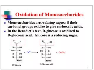
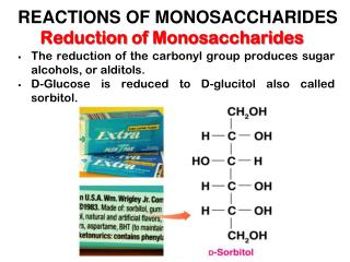
The Role of Pentose Phosphate Pathway (phosphogluconate pathway) (1) Synthesis ofNADPH(for reductive reactions in biosynthesis of fatty acids and steroids) (2) Synthesis ofRibose 5-phosphate (for the biosynthesis of ribonucleotides (RNA, DNA) and several cofactors) (3) Pentose phosphate pathway also provides a means for the metabolism of “unusual sugars”, 4, 5 and 7 carbons. Pentose phosphate pathway does not function in the production of high energy compounds like ATP.
Occurrence of the pentose phosphate pathway • Liver, mammary and adrenal glands, and adipose tissue • Red blood cells (NADPH maintains reduced iron) • NOT presentin skeletal muscles. • All enzymes in the cycle occur in the cytosol
Two phases: 1) The oxidative phase that generates NADPH 2) The nonoxidative phase (transketolase/ transaldolase system) that interconvert phosphorylated sugars.
Conversion of glucose-6-phosphate to 6-phosphogluconolactone
Conversion of 6-phosphogluconolactone to 6-phosphogluconate
Conversions of ribulose 5-phosphate Ribose 5-phosphate isomerase
Glucose + ATP + 2NADP+ + H2O ribose 5-phosphate + CO2 + 2NADPH + 2H+ + ADP The pentose phosphate pathway ends with these five reactions in some tissue. In others it continue in nonoxidative mode to make fructose 6-phosphate and glyceraldehyde 3-phosphate. These reactions link pentose phosphate pathway with glycolysis. The net reaction for the pentose phosphate pathway
Interconversions Catalyzed byTransketolase and Transaldolase • Transketolaseand transaldolase have broad substrate specificities • They catalyze the exchange of two- and three-carbon fragments between sugar phosphates • For both enzymes, one substrate is an aldose, one substrate is a ketose
Glucose-6-phosphate dehydrogenase deficiency NADPH is required for the proper action of the tripeptide glutathione (GSH)(maintains it in the reduced state). GSH in erythrocytes maintains hemoglobin in the reducedFe(II) state necessary for oxygen binding. GSH also functions to eliminate H2O2 and organic peroxides. Peroxides can cause irreversible damage to hemoglobin and destroy cell membranes.
Glucose-6-phosphate dehydrogenase deficiency – the most commonenzymopathy affecting hundreds of millions of people. About 10 % of individuals of African or Mediterranean descent have such genetic deficiency. Erythrocytes with a lowered level of reduced glutathione are more susceptible to hemolysis and are easily destroyed especially if they are stressed with drugs (for example, antimalarial drugs). In severe cases, the massive destruction of red blood cells causes death. Red blood cells with Heinz bodies. Dark particles (Heinz bodies) are denaturated proteins adhered to cell membranes.
Lactate • Glycolysis generates large amounts of lactate in active muscle • Red blood cells steadily produce lactate • Lactate produced by active skeletal muscle and erythrocytes is a source of energy for other organs • The plasma membranes of some cells, particularly cells in cardiac muscle, contain carriers that make them highly permeable to lactate and pyruvate. • Lactate and pyruvate diffuse out of active skeletal muscle into the blood and then into these permeable cells. • Once inside these well-oxygenated cells, lactate can be reverted back to pyruvate and metabolized through the citric acid cycle and oxidative phosphorylation to generate ATP. • The use of lactate in place of glucose by these cells makes more circulating glucose available to the active muscle cells. • Excess lactate enters the liver.
The Cori Cycle Liver lactate dehydrogenase converts lactate to pyruvate (a substrate for gluconeogensis) Glucose produced by liver is delivered to peripheral tissues via the bloodstream Contracting skeletal muscle supplies lactate to the liver, which uses it to synthesize glucose. These reactions constitute the Cori cycle