Download
1 / 32
340 likes | 846 Views
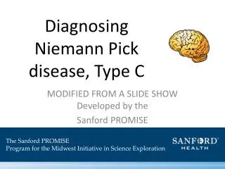
Diagnosing Niemann Pick disease, Type C. Developed by the Sanford PROMISE. The Sanford PROMISE Program for the Midwest Initiative in Science Exploration. Lab Safety. What’s wrong with this picture?. Lab Safety. What’s wrong with this picture? Gloves Goggles Lab coat Posture

E N D
Diagnosing Niemann Pick disease, Type C Developed by the Sanford PROMISE The Sanford PROMISE Program for the Midwest Initiative in Science Exploration
Lab Safety • What’s wrong with this picture?
Lab Safety • What’s wrong with this picture? • Gloves • Goggles • Lab coat • Posture • Work area • Shower/eyewash • Spills • Emergency exits
The Case Your summer job is as intern in a genetics lab at a Mount Blueberry Children’s hospital. A doctor comes to your team and says that he has a family in which he suspects three cousins of all have Niemann-Pick type C disease. The family would like to know: 1) the children indeed have Niemann-Pick type C 2) what are the risks of future children in the family developing the disease.
Niemann Pick Type C • Niemann-Pick disease is an inherited condition in which patients have abnormal lipid metabolism causing harmful amounts of lipids to accumulate in the spleen, liver, lungs, bone marrow, and brain. • Caused by mutations in genes NPC1, NPC2, SMPD1 • NPC1 mutations account for 95% of type C cases. Video of Lysosomal Storage Diseases
Micropipettes Push button/ Adjustable knob • What is a micropipette for? • Used for moving volumes of liquid from 0.2-1000μL • Adjustable/Fixed settings • Why should disposable micropipette tip be used? • To prevent sample and micropipette contamination Finger rest Tip ejector button Body Volume display Tip holder (shaft) Micropipette tip
Micropipette Setup • Setting the delivery volume • Pull out adjustment knob • Turn to adjust delivery volume • Check volume display while setting • Reading the volume display • Unique for each pipette • 20 – 200μL range 10μL 2 0 3 1 0 1
Micropipette Operation PRACTICE!
Part 1 – Polymerase Chain Reaction (PCR) • PCR is a technique used to amplify specific regions of DNA • Start with one molecule of double stranded patient DNA and generate 2 after one cycle • Exponential increase in DNA
Part 1 – Polymerase Chain Reaction (PCR) Step 1: Denature the double-stranded DNA into single strands. Step 2: Anneal the primers to a specific region of DNA. Step 3: Extend by synthesizing new DNA using the enzyme DNA polymerase which uses the original strand as a template for nucleotide placement.
Polymerase Chain Reaction (PCR) • What is in the PCR reaction mix? DNA Sample PCR Rxn Mix Thermocycler
Polymerase Chain Reaction (PCR) • What is in the PCR reaction mix? DNA Sample PCR Rxn Mix Thermocycler
Polymerase Chain Reaction • Step 1: Denature DNA • Heat it up! • Step 2: Primer annealing • Get the first tracks laid out • Step 3: Extension • DNA polymerase fills in the gaps
Polymerase Chain Reaction (PCR) • Cycling Conditions • Initial Denaturation • 95˚C for 2 minutes • Denaturation • 95˚C for 30 seconds • Primer Annealing • 60˚C for 20 seconds • Extension • 72˚C for 1 minute • Final Extension • 72˚C for 3 minutes 20 Cycles
Part 2 – Family History Different types of genetic mutations
Part 3 – DNA Electrophoresis • DNA electrophoresis is a technique used to separate DNA by charge and size • DNA is a charged molecule – what charge?
DNA Electrophoresis • DNA is separated on an agarosegel based on size • TAE buffer is added to cover the gel • A power supply applies a current across the gel
DNA Electrophoresis Cathode (negative) Anode (positive)
DNA Ladder • Where do we expect to see the DNA bands from our PCR reaction?
Hypothesis DNA Ladder Affected Unaffected Carrier 2000 bp 1500 bp 1000 bp 750 bp 500 bp 250 bp
DNA Electrophoresis • Place micropipette tip into TAE buffer directly over the well in the agarose gel • Slowly pipet sample into the well
DNA Visualization • DNA cannot be visualized with the visible eye • GelRed will bind to DNA • GelRed is in the agarose gel • GelRed is excited by UV light and will give off visible light ***Dangers of UV light***
Career Pathways Careers • DNA Scientist • Biomedical lab • Clinical lab • Forensic analysis • Paternity testing • Clinical Geneticist Regional Groups • Identity Genetics Inc. • Sanford Health