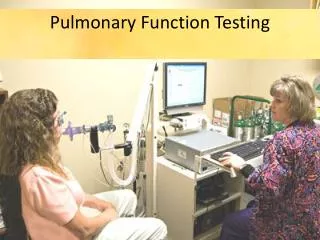
Pulmonary Function Testing
Pulmonary Function Testing. Dr. Alireza Safaiyan Occupational medicine specialist. PFT Pulmonary Function Testing. PFTs can include : Simple screening spirometry Formal lung volume measurement Diffusing capacity for carbon monoxide Arterial blood gases Inhalation challenge tests

Pulmonary Function Testing
E N D
Presentation Transcript
Pulmonary Function Testing Dr. AlirezaSafaiyan Occupational medicine specialist
PFTPulmonary Function Testing PFTs can include : • Simple screening spirometry • Formal lung volume measurement • Diffusing capacity for carbon monoxide • Arterial blood gases • Inhalation challenge tests • Exercise stress tests
Total lung capacity (TLC) FRC is usually measured by: • gas dilution technique or • body plethysmography
Gas dilution technique • Helium dilution techniques: based on the inhalation of a known concentration and volume of an inert tracer gas, such as helium, followed by equilibration of 7 to 10 minutes in the closed-circuit helium dilution technique. The final exhaled helium concentration is diluted in proportion to the unknown volume of air in the patient's chest (residual volume). • Nitrogen-washout technique: the patient breathes 100% oxygen, and all the nitrogen in the lungs is washed out. The exhaled volume and the nitrogen concentration in that volume are measured. The difference in nitrogen volume at the initial concentration and at the final exhaled concentration allows a calculation of intrathoracic volume, usually FRC.
Gas dilution technique • A limitation of this technique is that it does not measure air in noncommunicatingbullae, and therefore it can underestimate total lung capacity, especially in patients with severe emphysema.
Body plethysmography • principle of Boyle's law, which states that the volume of gas at a constant temperature varies inversely with the pressure applied to it • can measure the total volume of air in the chest, including gas trapped in bullae. • can be performed quickly
DLCODiffusing capacity of the lung for carbon monoxide • the single-breath breath-holding technique • a subject inhales a known volume of test gas that usually contains 10% helium, 0.3% carbon monoxide, 21% oxygen, and the remainder nitrogen. • holds his or her breath for 10 seconds. • exhales to wash out a conservative overestimate of mechanical and anatomic dead space. • an alveolar sample is collected. • DLCO is calculated from the total volume of the lung, breath-hold time, and the initial and final alveolar concentrations of carbon monoxide Hemoglobin concentration is a very important measurement in interpreting reductions in DLCO
Obstructive Lung Diseases Cystic fibrosis Emphysema Parenchymal Lung Disease Drug reactions (e.g., amiodarone, bleomycin) Idiopathic Interstitial lung disease Lung disease caused by fibrogenic dusts (e.g., asbestosis) Lung disease caused by biologic dusts (e.g., allergic alveolitis) Sarcoidosis Pulmonary Involvement in Systemic Diseases Dermatomyositis-polymyositis Inflammatory bowel disease Mixed connective tissue disease Progressive systemic sclerosis Rheumatoid arthritis Systemic lupus erythematosus Wegener's granulomatosis Cardiovascular Diseases Acute and recurrent pulmonary thromboembolism Acute myocardial infarction Fat embolization Mitral stenosis Primary pulmonary hypertension Pulmonary edema Other Acute and chronic ethanol ingestion Bronchiolitisobliterans with organizing pneumonia (BOOP) Chronic hemodialysis Chronic renal failure Ciagarette smoking Cocaine freebasing Diseases associated with anemia Marijuana smoking Increases In DLCO Diseases associated with increased pulmonary blood flow (e.g., left-to-right intracardiac shunts) Diseases associated with polycythemia Exercise Pulmonary hemorrhage
Exhaled Nitric Oxide • The measurement of exhaled nitric oxide as a reflection of airway inflammation is gaining rapid acceptance as a pulmonary function test
Inhalation challenge tests (Bronchoprovocation) • To define nonspecific airway hyperreactivity • Methacholineand histamine are the agents most often used with this procedure inhaled through a nebulizer, although other agents may also be useful. • a five-stage procedure with five different increasing concentrations.(0.0625 – 16 mg/ml) After each stage, the patient performs a spirometry. • a 20% reduction in the FEV1 (PC20FEV1 ) is significant. • In rare cases, a bronchospasm can occur with inhalation challenge testing.
Exercise stress tests • Exercise stress tests evaluate the effect of exercise on lung function tests. Spirometry readings are done after exercise and then again at rest. • Six-minute walk test
Definition of spirometry • A physiological test for measuring volumes inhaled or exhaled by an individual as a function of time • A non-invasive method of evaluation of pulmonary function • Not for definitive diagnosis • Simple, cost effectiveness, accessible • Needs patient cooperation
Indication • Not a screening test for general population • Diagnostic • Monitoring • Impairment evaluation • Public health
Diagnostic To evaluate symptoms • Chest pain • Cough • Dyspnea • Orthopnea • Phlegm production To evaluate signs • Wheezing • Chest deformity • Cyanosis • Diminished breath sounds • Expiratory slowing • Overinflation • Unexplained crackles To evaluate abnormal laboratory tests • Abnormal chest radiographs • Hypercapnia • Hypoxemia • Polycythemia To measure the effect of disease on pulmonary function To screen persons at risk for pulmonary diseases • Smokers • Persons in occupations with exposures to injurious substances Some routine physical examinations • To assess preoperative risk • To assess prognosis (lung transplant, etc.) • To assess health status before enrollment in strenuous physical activity programs
Monitoring To assess therapeutic interventions • Bronchodilator therapy • Steroid treatment for asthma, interstitial lung disease, etc. • Management of congestive heart failure • Other (antibiotics in cystic fibrosis, etc.) To describe the course of diseases affecting lung function • Pulmonary diseases • Obstructive small airway diseases • Interstitial lung diseases • Cardiac diseases • Congestive heart failure • Neuromuscular diseases • Guillain-Barrésyndrome • To monitor persons in occupations with exposure to injurious agents To monitor for adverse reactions to drugs with known pulmonary toxicity
Evaluation of Disability or Impairment To assess patients as part of a rehabilitation program • Medical • Industrial • Vocational To assess risks as part of an insurance evaluation To assess persons for legal reasons • Social Security or other government compensation programs • Personal injury lawsuits • Other
Public Health • Epidemiologic surveys • Comparison of health status of populations living in different environments • Validation of subjective complaints in occupational or environmental settings • Derivation of reference equations
Indications in occupational medicine • Primary prevention(Pre-employment) • physical demands of a job require a certain level of cardiopulmonary fitness, eg, heavy manual labor or firefighting • Respirator use can impose a significant burden on the cardiopulmonary systems, eg, use of a self-contained breathing apparatus, or prolonged use of certain negative-pressure masks under conditions of heavy physical exertion and/or heat stress • Research (Respiratory hazards)
Secondary prevention Medical surveillance programs & periodic evaluation OSHA : • asbestos, cadmium, coke oven emissions, or cotton dust • respirator-wearers exposed to benzene, formaldehyde methylene chloride • Silicosis • Tertiary prevention • Follow-up spirometry • Workers’ compensation setting
Contraindications • Active hemoptysis • Pneumothorax • Unstable Cardiovascular status (6 w) • Cerebral/Thoracic/Abdominal aneurysm • Recent eye surgery • Acute disorder that may interfere with performance (e.g, vomiting) • Thoracic or abdominal surgery( 3 w) • Recent CVA or pulmonary emboli • Respiratory distress
Confounding factors • Common cold (previous 3 days) • Severe respiratory infection (previous 3w) • Smoking( 1hr) • Heavy food (1hr) • Bronchodilator use
Complications • Chest pain • Syncope, dizziness • Increased ICP • Paroxysmal coughing • Nosocomial infection • Bronchospasm
Type of spirometer Flow-type spirometer Volumetric spirometer
Hygiene & infection control • Hand washing • Gloves • Disposable mouth piece & nose clip • Disinfection or sterilization of reusable mouth piece • Extra precautions for patient with known transmissible infection
Normality and predicted equations • lung function, such as FEV1 or FVC, are affected most significantly by standing height, age, gender, race, and, to a lesser extent, weight • These reference standards are based on a cohort of normal subjects of similar age, height, and race, withnormal being defined as persons without a history of smoking or disease that can affect lung function.
Reference values • Knudson (male/ female) • National Health and Nutrition Examination Survey (NHANES III) • ERS (ECCS) • ATS • Golshan • ITS
Spirometric values FVC—Forced vital capacity; the total volume of air that can be exhaled during a maximal forced expiration effort. FEV1—Forced expiratory volume in one second; the volume of air exhaled in the first second under force after a maximal inhalation. FEV1/ FVC ratio—The percentage of the FVC expired in one second. FEV6 —Forced expiratory volume in six seconds. FEF25–75%—Forced expiratory flow over the middle one half of the FVC; the average flow from the point at which 25 percent of the FVC has been exhaled to the point at which 75 percent of the FVC has been exhaled. MVV—Maximal voluntary ventilation. This measures the greatest amount of air you can breathe in and out during one minute. Lung volumes ERV—Expiratory reserve volume; the maximal volume of air exhaled from end-expiration. IRV—Inspiratory reserve volume; the maximal volume of air inhaled from end-inspiration. RV—Residual volume; the volume of air remaining in the lungs after a maximal exhalation. VT —Tidal volume; the volume of air inhaled or exhaled during each respiratory cycle. Lung capacities FRC—Functional residual capacity; the volume of air in the lungs at resting end-expiration. IC—Inspiratory capacity; the maximal volume of air that can be inhaled from the resting expiratory level. TLC—Total lung capacity; the volume of air in the lungs at maximal inflation. VC—Vital capacity; the largest volume measured on complete exhalation after full inspiration.
کاليبراسيون دستگاه • کاليبراسيون روزانه يا هر 4 ساعت در موارد شلوغ • سرنگ مخصوص کاليبراسيون(1 ليتري يا 3 ليتري) • خطاي پذيرفته شده 3% يا 90 سي سي است(2/91-3/09)
Subject maneuvers • FVC maneuver • Closed circuit • Open circuit • Well-fitting false teeth → yes or no • Sitting or standing • Nose clip • Procedure 1. Inhale compete 2. Exhale: with minimal hesitation “blast” not just “blow” “keep going”
maneuver evaluation • Start of test criteria - Extrapolation volume(EV < 5% of FVC or150 ml) -Time-to-PEF < 0.120 s • End of test criteria - the subject cannot or should not continue - exhalation at least 6s (in children <10 yrs: at least 3s) - volume-time curve show no change in volume (<0.025 lit) for at least 1s • In obstruction or older subjects more than 6s exhalation (till 15s)
Acceptability • Start of test criteria • End of test criteria • Cough especially during first second • Valsalva maneuver (glottis closure) • Leak from the mouth • Obstruction of the mouthpiece • Extra breath during the maneuver • At the most eight tests should be performed
Reproducibility • At least three acceptable maneuvers Maximum difference between the largest and next largest FVC andFEV1 = 150ml or 5% (If FVC <1lit, this value is 100ml)