Bone
Bone. Gross histology Bone matrix and bone cells (osteoblast, osteocyte and osteoclast) Regulation of osteoblast/osteocyte/osteoclast formation Endochondral and intramembranous bone formation Bone remodeling. Development and Maintenance of the Skeleton.

Bone
E N D
Presentation Transcript
Bone • Gross histology • Bone matrix and bone cells (osteoblast, osteocyte and osteoclast) • Regulation of osteoblast/osteocyte/osteoclast formation • Endochondral and intramembranous bone formation • Bone remodeling
Development and Maintenance of the Skeleton Development Maintenance Osteoporosis Age 0-20 20-50 50+ Sequence BF BR BR BF BR BF Modeling Remodeling Activity BF > BR BF = BR BF < BR Uncoupled Coupled Uncoupled Marks S.C. and Hermey, D.C. The Structure and Development of Bone; Principles of Bone Biology, Blezikian, Raisz and Rodan, eds, 1996.
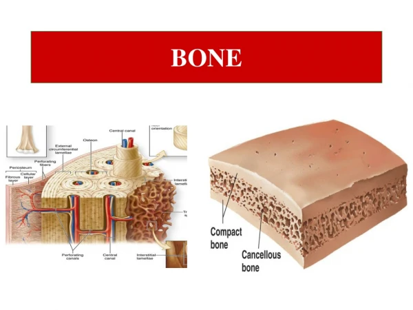
Flat (skull and pelvis) and long (femur, tibia) bones Two types of bone macroscopically: 1. Compact bone: dense outer sheet covering flat bones and shaft of long bones; mature bone 2. Trabecular/cancellous/spongy bone: present within the central marrow cavity comprised of network of delicate bars and sheet of trabecular bone which branch and intersect to form a sponge-like network Periosteum: layer of dense connective tissue surrounding bone. Has 2 layers: Outer fibrous layer and inner layer next to bone containing bone cells their precursors and blood vessels Endosteum: layer if thin connective tissue lining the inner surface of bone facing bone marrow
Microscopic appearance (both compact and trabecular) Arranged as lamellae (microscopic layer) 3 distinct lamellae: circumferential concentric and interstitial concentric Concentric lamellae forms the basic metabolic unit of bone the osteon (haversian system)
Bone Matrix Bone Matrix is physiologically mineralized and is constantly regenerated throughout life as a consequence of bone turnover Bone consists various bone matrix proteins that play an integral part in bone function The predominant and basic building block of bone is type I collagen However, trace amounts of type III and V collagens are also present
Type I Collagen Most abundant protein in bone matrix (~ 85 - 90% of organic matrix) Provides elasticity, flexibility and serves as a scaffold determining shape Binds and orients other proteins that nucleate hydroxyapatite deposition Collagen undergo several post translational modifications (hydroxylation of certain lysyl and hydroxylysyl residues), which are measured in urine as a parameter for bone resorption The animal model with defective type I collagen production has a condition similar to osteogenesis imperfecta, where the bones are mechanically weak and mineral crystals small
Type I collagen 19 types of collagen known so far in humans. Types 1, 2, 3 and 4 are most common Predominantly synthesized by fibroblasts Rod-like protein. Type I collagen has a length of ~ 300 nm, 1.5 nm in diameter and consists of a right handed triple helix of subunits composed of two 1(1) chain and one 2(1) chain There are 3 amino acids per turn and every 3rd amino acid is a G Synthesized as type 1 procollagen that contain an additional 150 amino acids at the N-terminal and 250 at the c-terminal
Normal OI
The Origin and Location of Bone Cells Marks S.C. and Hermey, D.C. The Structure and Development of Bone; Principles of Bone Biology, Blezikian, Raisz and Rodan, eds, 1996.
Osteoprogenitor cells: Contribute to maintaining the osteoblast population and bone mass. Located in the periosteum and endosteum and differentiate into osteoblasts Osteoblasts: synthesize the bone matrix on the bone forming surfaces Osteocytes: organized throughout the mineralized bone matrix that support bone architecture Osteoclasts: large multinucleated cells derived from fusion of monocytes/macrophages and resorbs bone.
Osteoblasts are located on the periosteal and endosteal bone surfaces and are responsible for bone formation through secretion of the organic components of bone matrix
Functions of Osteoblasts • 1. Main Function of osteoblast: Secretion of a complex • mixture of bone matrix proteins (osteoid) • Secretion of osteoid is unidirectional towards the bone surface • Osteoblasts are normally separated form the mineralized bone • matrix by a thin layer of unmineralized matrix (osteoid seam) • Regulate differentiation and activity of osteoclasts • Indirectly maintain calcium homeostasis • Indirectly responsible for mineralization of osteoid
Origin of Osteoblasts Limited self renewal osteoblasts Unlimited self renewal Runx2, Osx Runx2 Osx PPAR2 PPAR2 adipocytes Sox9 Multipotential Daughter Cell chondroblasts Tri-Bipotential Progenitor Cell MyoD myoblasts Stem Cell fibroblasts Decreasing proliferation Increasing differentiation Adapted from Primer on the metabolic bone diseases and disorders of mineral metabolism. Lian JB, Stein GS and Aubin JE., Bone formation: Maturation and functional activities of osteoblast lineage cells, ASBMR, 2003
Runx2 is a bone-related transcription factor that is essential for osteoblast differentiation and bone formation Wildtype Runx2-/- Otto et al., 1997
Osteoprogenitor Cells: Mesenchymal cells located on periosteal surfaces and bone marrow stromal cells are a good source of osteoblasts which act in concert with osteoclasts to model bone during growth and maintain bone architecture during adulthood Commitment of mesenchymal stem cells (MSCs) to tissue-specific types is mediated by trancriptional regulators that serve as “master switches” BMP2/4/7 induce transcription factors that mediate commitment of early progenitors (stem cells) toward a osteoblast phenotype
Pre-osteoblast > Osteoblast > Pre-osteocyte > Osteocyte Proliferation Matrix Maturation Mineralization Apoptosis Collagen Alk Phos Fra-2/JunD Cbfa1 Dlx-5 MGP Histone Collagen TGF Msx-2 cFos/cJun Id, Twist Osteopontin Bone Sialo- Protein(BSP) Osteocalcin Collagenase Bax Growth Differentiation Proliferation > < Mineralization + ECM In Vitro Development of Osteoblast Phenotype
The osteoblast developmental sequence: Lessons from cell culture studies • 3 stages of bone cell differentiation are recognized according to the proteins and • mRNA expressed at different stages: • Proliferation and extracellular matrix (ECM) biosynthesis • ECM development, maturation and organization • ECM mineralization
Osteoblast Differentiation • Proliferation and ECM biosynthesis: Increased mitotic activity with expression of • cell cycle and cell growth regulated genes that support proliferation • During this proliferation period, and fundamental to development of bone phenotype, • several genes associated with formation of the ECM (type I collagen, fibronectin, • and transforming growth factor- (TGF- ) are actively expressed and then gradually • down-regulated with collagen mRNA being maintained at low basal level during • subsequent stages of osteoblast differentiation • Immediately after down-regulation of proliferation, proteins associated with the bone • cell phenotype are detected • Alkaline phosphatase levels increase 10-fold in the immediate post-proliferative phase • With the onset of mineralization, other bone-related genes are induced: bone • sialoprotein (BSP), osteopontin and osteocalcin are increased which parallels • mineral deposition
Osteocytes: Terminally differentiated osteoblasts Osteocytes: terminally differentiated osteoblasts that support bone structure and metabolic function Osteocytes develop by forming numerous cytoplasmic connections with adjacent cells to ensure their viablity as the mineralizing osteoid renders the ECM impermeable Osteocytes are present in lacunae and has numerous cellular extensions of filapodial processes Osteocytes express OCN, galectin-3 and CD44 (a cell adhesion receptor for hyaluronate and several bone matrix proteins that are involved in cellular extensions) Osteocytes are continuous with the lining osteoblasts and is necessary for transmitting mechanosensory signals such as transducing stress signals (streching, bending) to biological activity
Multinucleated cell the originates from hematopoietic stem cells Characterized by possessing tartrate-resistant acid phosphatase (TRAP) within its cytoplasmic vesicles and vacuoles Found against bone surface in hollowed depressions called Howship’s lacunae RANK: receptor-activated nuclear factor κB and RANKL: RANK ligand
Ruffled border: Adjacent to resorbing bone surface the osteoclast is closely apposed to bone and has deep folds called ruffled border. Adjacent cytoplasm does not have any organelles and is enriched in actin. This zone is called the clear or sealing Zone. This enables the osteoclast to attach to mineralized surface and creates an acidic microenvironment which demineralizes bone and exposes the organic matrix. The matrix is degraded by enzyme acid phosphatase and cathepsin B
Bone Formation Endochondral bone formation
Bone Remodeling Five Phases of Remodeling 1. Activation of Osteoclasts 2. Resorption of Bone 3. Reversal Phase 4. Formation of Bone Activation of Osteoblasts Mineralization 5. Resting
Mineralization Physiologic bone mineralization in mammals refers to the ordered deposition of apatite on a type I collagen matrix The mineral is an analog of the geologic material, hydroxyapatite [Ca10(PO4)6(OH)2] - provides mechanical rigidity and load bearing strength to the bone composite Bone mineral also contains numerous impurities (carbonate, magnesium, acid phosphate) and vacancies (missing OH-) and is usually referred to as a poor, crystalline, carbonate-substituted apatite. These small imperfect crystals are more soluble that geologic apatite, enabling bone to act as a reservoir for calcium, phosphate, and magnesium ions
Steps in Mineralization: 1. Initiation 2. Propagation 3. ECM mineralization Bone Mineral is deposited at discrete sites in collagenous matrix As bone matures, the mineral crystals become larger and more perfect (containing fewer impurities) The increase in crystal dimension is due both to the actual addition of ions to the crystals (crystal growth) and to aggregation of the crystals
Initiation Not dependent on alkaline phosphatase Propagation ECM mineralization Dependent on alkaline phosphatase